

Development and preliminary evaluation of a novel preoperative index for quantitative analysis of photoreceptor loss in full-thickness macular holes
- PMID: 39460786
- PMCID: PMC11953196
- DOI: 10.1007/s00417-024-06654-z
Development and preliminary evaluation of a novel preoperative index for quantitative analysis of photoreceptor loss in full-thickness macular holes
Abstract
Purpose: To identify novel quantitative parameters for evaluating photoreceptor loss in full-thickness macular holes (FTMH), exploring their potential clinical impact on postoperative functional and anatomical recovery.
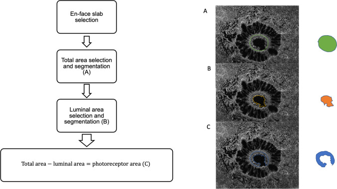
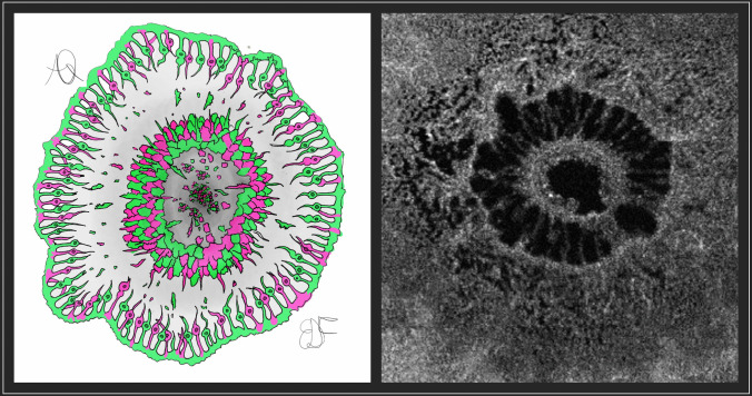
Methods: This pilot study enrolled 38 eyes from 38 patients diagnosed with FTMH. Preoperatively, eyes underwent analysis and were subsequently followed for six months post-surgery. Best-corrected visual acuity (BCVA) was recorded, and cross-sectional images of FTMH were obtained using B-scan optical coherence tomography (OCT) and en-face OCT. Quantitative assessment of ellipsoid zone (EZ) and external limiting membrane (ELM) integrity changes was conducted and correlated with postoperative anatomical and functional recovery. The photoreceptor Integrity Index (PIIN), calculated as the ratio of photoreceptor area to lumen hole area measured at customized segmentation, was correlated with the minimum and base diameters of the hole, positive change in BCVA, preoperative EZ defect (EZd), preoperative ELM defect (ELMd), and changes in EZ and ELM over the six-month follow-up period (∆-EZ and ∆-ELM). The main outcome measures focused on evaluating the effectiveness of PIIN in predicting postoperative anatomical and functional changes.
Results: A higher PIIN correlated with a greater BCVA change over six months (p < 0.001). Univariate regression analysis using the PIIN as a predictor for positive change in BCVA (|∆-BCVA| [logMAR]) over time yielded significant results (p < 0.001). Additionally, the PIIN significantly correlated with EZd at baseline, ELM at baseline, and ELMd change over the six-month follow-up period.
Conclusion: The PIIN shows promise as a tool for evaluating photoreceptor loss in macular holes and estimating postoperative functional and anatomical recovery.
Key messages: What is known Previous studies have extensively used optical coherence tomography (OCT) to investigate various biomarkers for assessing patients with full-thickness macular hole (FTMH), without considering detailed MH ultrastructural features Existing indexes used to predict surgical outcomes for FTMH primarily depend on geometrical parameters and do not integrate detailed ultrastructural characteristics, such as cellular components. What is new A novel concept introduces the quantitative measurement of residual photoreceptors located at the edge of FTMH. The Photoreceptor Integrity Index (PIIN) integrates different ultrastructural components of macular holes, aiming to become a valuable clinical tool to predict both anatomical and functional recovery outcomes following surgical intervention for FTMH.
Keywords: Full-thickness macular hole; Macular hole borders; PIIN; Photoreceptors.
© 2024. The Author(s).
Conflict of interest statement
Declarations. Financial disclosures: No financial disclosures. Informed consent: All research and measurements adhered to the tenets of the Declaration of Helsinki and were approved by the Institutional Review Board of University G. D’Annunzio of Chieti-Pescara [IRB NUMBER OMH-1055]. Informed consent was obtained from each patient after a detailed explanation of the nature and possible consequences of the study procedures. Competing interests: The authors declare that they have no competing interests.
Figures


References
-
- Ruiz-Moreno JM, Staicu C, Piñero DP, Montero J, Lugo F, Amat P (2008) Optical coherence tomography predictive factors for macular hole surgery outcome. Br J Ophthalmol. 92(5):640–644 - PubMed
-
- Matet A, Savastano MC, Rispoli M et al (2015) En face optical coherence tomography of foveal microstructure in full-thickness macular hole: a model to study perifoveal Müller cells. Am J Ophthalmol. 159(6):1142-1151.e3 - PubMed
-
- Iwasaki M, Ando R, Aoki S, Miyamoto H (2022) Restoration process of the outer retinal layers after surgical macular hole closure. Retina. 42(2):313–320 - PubMed
MeSH terms
LinkOut - more resources
Full Text Sources
Research Materials
