. 2024 Dec 20;5(4):103417.
doi: 10.1016/j.xpro.2024.103417. Epub 2024 Oct 25.

Protocol to profile spatially resolved NLRP3 inflammasome complexes using APEX2-based proximity labeling

Affiliations

Protocol to profile spatially resolved NLRP3 inflammasome complexes using APEX2-based proximity labeling

Zhu Liang et al. STAR Protoc. .

Abstract

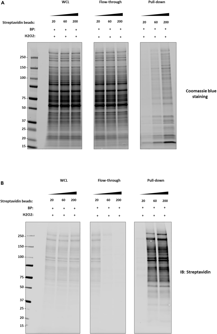
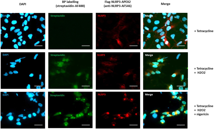
The NLRP3 inflammasome is a key multi-protein complex controlling inflammation, particularly interleukin-1β (IL-1β) production. Here, we present a protocol to profile spatially resolved NLRP3 inflammasome complexes using ascorbic peroxidase 2 (APEX2)-based proximity labeling combined with liquid chromatography-tandem mass spectrometry (LC-MS/MS). We describe steps for design and generation of the fusion construct, characterization of the stable FLAG-NLRP3-APEX2 expression cell line by western blotting/imaging, biotinylated proteome enrichment, and mass spectrometry analysis. For complete details on the use and execution of this protocol, please refer to Liang et al.1.

Keywords: immunology; mass spectrometry; molecular biology; proteomics.


Conflict of interest statement

Declaration of interests The authors declare no competing interests.

Figures

None
Graphical abstract
Figure 1
Figure 1
Schematic representation of APEX2 proximity labeling (A) Once activated, APEX2 catalyzes the oxidation of a biotin-phenol substrate, generating highly reactive biotin-phenoxyl radicals. (B) The biotin-phenoxyl radicals quickly interact with nearby electron-rich amino acids, particularly tyrosine (tryptophan and cysteine in some cases). The radical reacts with the aromatic ring of tyrosine through a radical coupling mechanism, resulting in biotinylation of the protein.
Figure 2
Figure 2
Optimization of the streptavidin beads for the enrichment of biotinylated proteins (A and B) Coomassie blue staining (A) and streptavidin blotting (B) of whole cell lysates (WCL), flow-through, and pull-down fractions after streptavidin bead enrichment. Different amounts of streptavidin beads (20, 60, 200 μL) were used for the pull-down experiments as indicated.
Figure 3
Figure 3
Capture and biotinylation of ASC in HEK293 cells stably expressing Flag-NLRP3-APEX2 (A) Schematic overview of the experimental design for treatment conditions and APEX2 induced biotinylation. Flp-In T-REx HEK293 cell lines stably expressing flag-NLRP3-APEX2 were treated with either DMSO or Nigericin (10 μM) for 90 min, followed by incubation with 500 μM biotin-phenol (BP) and 1 mM H2O2 for 20 s. The diagram illustrates the biotinylation process and the potential interaction with ASC protein under different treatment conditions. (B and C) Streptavidin blotting of whole cell lysates (B) and Immunoblotting of the streptavidin beads-pulled down samples (C) from HEK293 cells stably expressing flag-NLRP3-APEX2 under various conditions as indicated. Figure reprinted with permission from Liang et al.
Figure 4
Figure 4
APEX2 labeling is able to capture the formation of NLRP3 puncta upon activation Confocal fluorescence imaging of NLRP3 APEX2 labeling was conducted in HEK293 cell lines. NLRP3-APEX2 HEK293 stable cell lines were treated with tetracycline 16 h, followed by a 90-min treatment with either Nigericin (10 μM) or DMSO (as a control). Subsequently, the cells were incubated with biotin phenol and then H2O2, as specified. After incubation, the cells were fixed and stained with a streptavidin-Alexa Fluor 488 (AF488) conjugate to visualize biotinylated proteins and with an anti-NLRP3 antibody to determine the localization of NLRP3-APEX2. Biotinylated proteins (BP) were visualized, and scale bars represent 50 μm. Figure reprinted with permission from Liang et al.
Figure 5
Figure 5
Characterization of the NLRP3-APEX2 fusion construct by western blotting (A) Schematics of the experimental workflow. Cells are lysed in RIPA buffer and cleared by centrifugation. Biotinylated proteins are captured using streptavidin beads, followed by washes with RIPA, KCl, Na2CO3, and 2 M Urea before being resuspended in RIPA. Enriched biotinylated proteins are either digested on beads with trypsin or eluted by boiling. (B–D) Western blotting of whole cell lysate (B), streptavidin-captured proteins (C) and flow-through (D) using the antibodies as indicated. Figure reprinted with permission from Liang et al.
Figure 6
Figure 6
High background in the negative control during streptavidin pull-down Streptavidin blotting (left) and Coomassie blue staining (right) of the pull-down fraction after streptavidin bead enrichment from an APEX2 labeling.

References

    1. Liang Z., Damianou A., Vendrell I., Jenkins E., Lassen F.H., Washer S.J., Grigoriou A., Liu G., Yi G., Lou H., et al. Proximity proteomics reveals UCH-L1 as an essential regulator of NLRP3-mediated IL-1beta production in human macrophages and microglia. Cell Rep. 2024;43 doi: 10.1016/j.celrep.2024.114152. - DOI - PubMed
    1. Burckstummer T., Bennett K.L., Preradovic A., Schutze G., Hantschel O., Superti-Furga G., Bauch A. An efficient tandem affinity purification procedure for interaction proteomics in mammalian cells. Nat. Methods. 2006;3:1013–1019. doi: 10.1038/nmeth968. - DOI - PubMed
    1. Gregan J., Riedel C.G., Petronczki M., Cipak L., Rumpf C., Poser I., Buchholz F., Mechtler K., Nasmyth K. Tandem affinity purification of functional TAP-tagged proteins from human cells. Nat. Protoc. 2007;2:1145–1151. doi: 10.1038/nprot.2007.172. - DOI - PMC - PubMed
    1. S N.P., Kwon K. The HaloTag: Improving Soluble Expression and Applications in Protein Functional Analysis. Curr Chem Genomics. 2012;6:8–17. doi: 10.2174/1875397301206010008. - DOI - PMC - PubMed
    1. Stagge F., Mitronova G.Y., Belov V.N., Wurm C.A., Jakobs S. SNAP-CLIP- and Halo-tag labelling of budding yeast cells. PLoS One. 2013;8 doi: 10.1371/journal.pone.0078745. - DOI - PMC - PubMed

Publication types

MeSH terms

LinkOut - more resources