. 2024 Oct 26;14(1):25555.
doi: 10.1038/s41598-024-76482-y.

The Tmem16a chloride channel is required for mucin maturation after secretion from goblet-like cells in the Xenopus tropicalis tadpole skin

Affiliations

The Tmem16a chloride channel is required for mucin maturation after secretion from goblet-like cells in the Xenopus tropicalis tadpole skin

Eamon Dubaissi et al. Sci Rep. .

Abstract

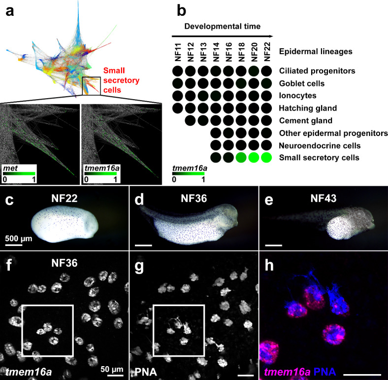
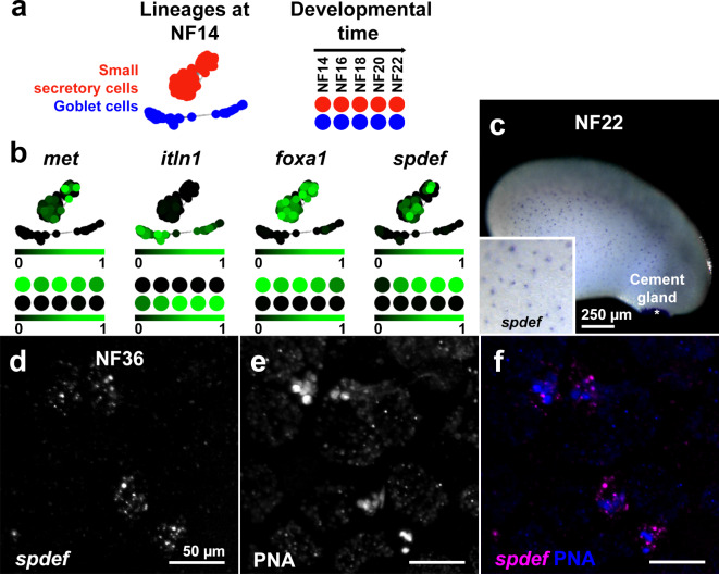
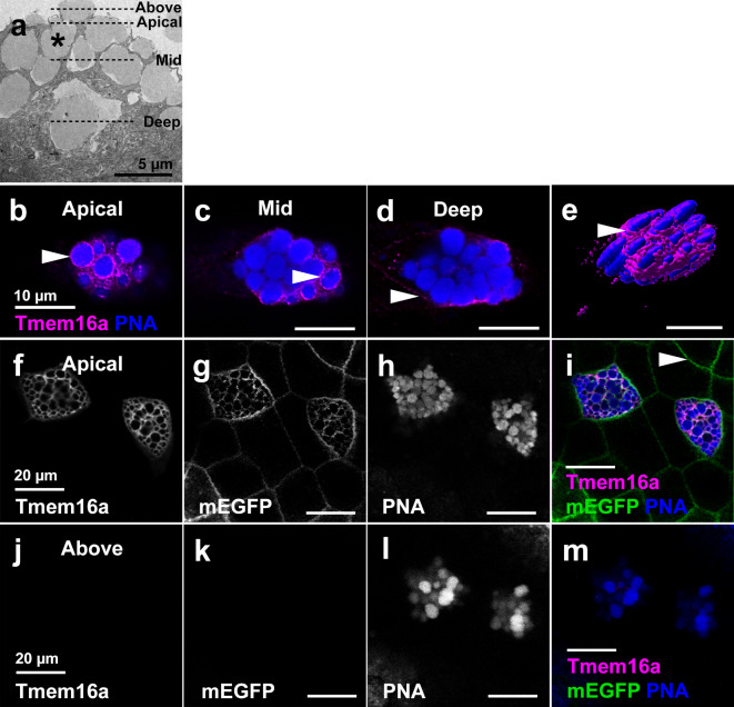
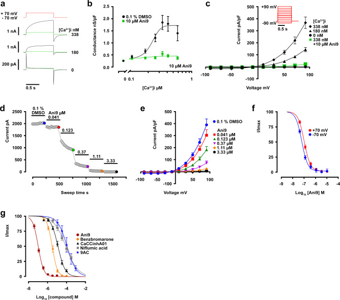
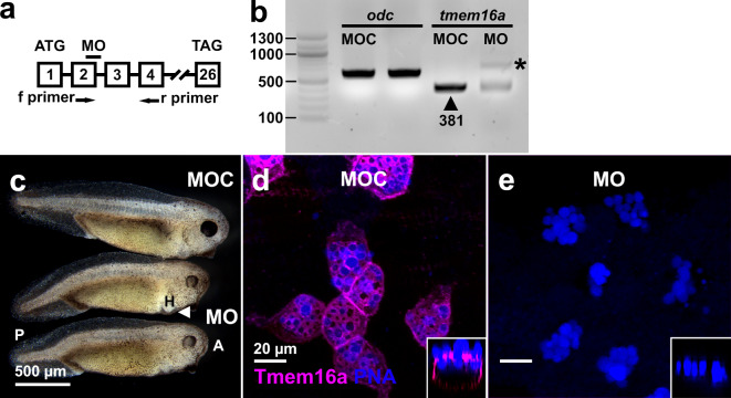
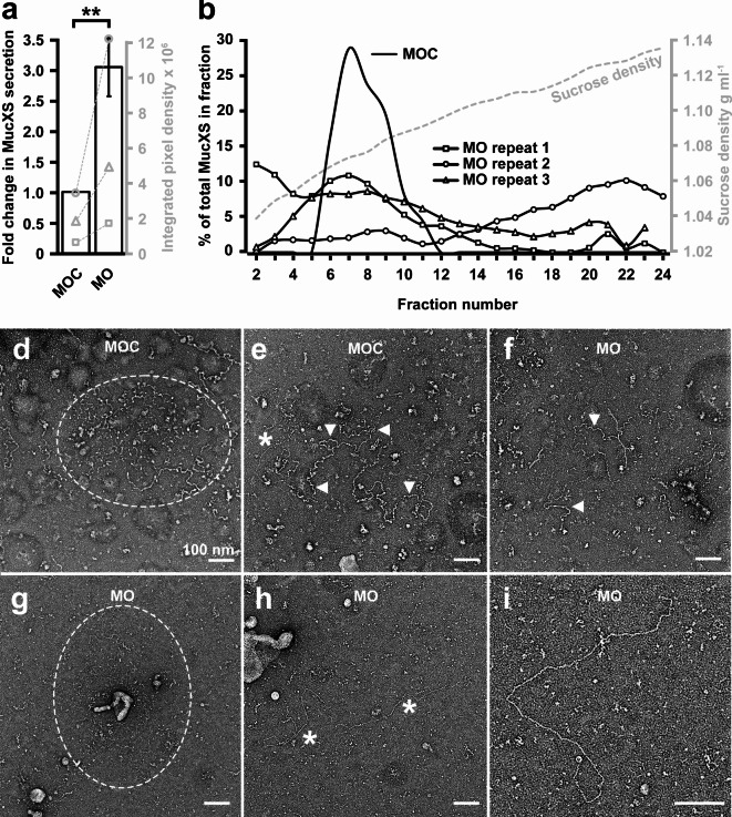
The TMEM16A chloride channel is proposed as a therapeutic target in cystic fibrosis, where activation of this ion channel might restore airway surface hydration and mitigate respiratory symptoms. While TMEM16A is associated with increased mucin production under stimulated or pro-inflammatory conditions, its role in baseline mucin production, secretion and/or maturation is less well understood. Here, we use the Xenopus tadpole skin mucociliary surface as a model of human upper airway epithelium to study Tmem16a function in mucus production. We found that Xenopus tropicalis Tmem16a is present at the apical membrane surface of tadpole skin small secretory cells that express canonical markers of mammalian "goblet cells" such as Foxa1 and spdef. X. tropicalis Tmem16a functions as a voltage-gated, calcium-activated chloride channel when transfected into mammalian cells in culture. Depletion of Tmem16a from the tadpole skin results in dysregulated mucin maturation post-secretion, with secreted mucins having a disrupted molecular size distribution and altered morphology assessed by sucrose gradient centrifugation and electron microscopy, respectively. Our results show that in the Xenopus tadpole skin, Tmem16a is necessary for normal mucus barrier formation and demonstrate the utility of this model system to discover new biology relevant to human mucosal biology in health and disease.

Keywords: Xenopus tropicalis; Ion channel; Mucin; Mucus; TMEM16A.


Conflict of interest statement

HD and MG are employees and stockholders of Enterprise Therapeutics, which has previously developed TMEM16A modulators. ED, ENH, SL, RC, Ch, PM, RKG, ISR and DJT declare no competing interests.

Figures

Fig. 1
Fig. 1
X. tropicalis tmem16a is expressed in SSCs in the tadpole skin. (ab) In a developmental time series of single-cell transcriptomes in X. tropicalis embryos from NF8-NF22, an SSC lineage (highlighted in red) was identified (a, upper). An expanded view (black box) examining gene-specific expression in this SSC lineage reveals expression of met (a, lower left; marking this SSC lineage) and of tmem16a (a, lower right). Within epidermal lineages, tmem16a expression is present in the SSC lineage from NF18 but absent from other epidermal lineages from differentiation to NF22 (b). Each circle represents a developmental stage in a specific epidermal lineage. Scale bars: colour intensity represents normalised expression level from undetectable (0) to maximum expression (1) within the entire dataset. (ce) Chromogenic in situ hybridisation for tmem16a at stages NF22 (c), NF36 (d) and NF43 (e) reveals punctate expression throughout the skin, matching typical SSC distribution. (fh) Fluorescent in situ hybridisation for tmem16a (f) and dual staining with the SSC vesicle marker PNA (g) confirms that tmem16a is expressed in SSCs (merged and expanded from white box in h).
Fig. 2
Fig. 2
SSCs are a discrete epidermal cell type that express classical airway GC markers. (ab) SSC (red) and GC (blue) lineage clusters in X. tropicalis embryos at NF14 and from NF14-22, are discrete (a) and marked by lineage-specific expression of met and itln1, respectively (b). The canonical goblet cell markers, foxa1 and spdef, are expressed almost exclusively in the SSC cell cluster at NF14, and over developmental time (b). Scale bars: colour intensity represents normalised expression level from undetectable (0) to maximum (1) within the dataset. c. Chromogenic in situ hybridisation for spdef at NF22 reveals punctate expression throughout the skin (inset), typical of SSC distribution, and strong expression in the cement gland (asterisk). (df) Fluorescent in situ hybridisation of spdef mRNA (d) and dual staining with PNA (e) confirms that spdef is expressed in SSCs (merged in f).
Fig. 3
Fig. 3
Tmem16a localises to the plasma and not vesicle membrane in SSCs. (a) SEM of SSCs reveals the presence of large vesicles (example indicated by asterisk) that bulge beyond the apical cell membrane of SSCs. Dashed lines indicate the planes of view in (bm). (be) Immunofluorescent localisation of Tmem16a protein in apical, mid and basal planes of a single SSC reveals Tmem16a is present in a pattern that, in the apical (b) and mid (c) planes, appears to surround the PNA-positive secretory vesicles regions of cell (arrowheads). However, this localisation pattern is absent around vesicles in the more basal region of the cell (d), and expression becomes evident in the presumed plasma membrane (d, arrowhead). 3D surface rendering shows Tmem16a at an apical plane below that of the bulging vesicles (e). (f-i) In the apical plane of epidermal cells, Tmem16a (f) localises with mEGFP (g) in SSCs stained with PNA (h; merged in i), but does not localise with mEGFP marking the plasma membrane of other cell types (h, arrowhead). (jm) Above the plane of epidermal cells, Tmem16a (j) and mEGFP (k) immunofluorescence is absent from bulging PNA-positive secretory vesicles (l; merged in m), demonstrating that Tmem16a is absent from the secretory vesicle membrane.
Fig. 4
Fig. 4
Biophysical and pharmacological characterisation of X. tropicalis Tmem16a. (a) Example currents evoked by single depolarizing pulses from − 70 to + 70 mV when [Ca2+]i is 0, 180 (Ca2+ EC20) or 338 nM (Ca2+ EC100), with inhibition in the presence of 10 μm Ani9 shown in green. (bX. tropicalis Tmem16a conductance is dependent on intracellular calcium levels (black) and inhibited in the presence of Ani9 (green). (c) In whole-cell current-voltage (I-V) tests recorded at 0, 180 & 338 nM [Ca2+]i, the inhibitory effect of Ani9 on maximally-active current (at 338 nM [Ca2+]i) across the voltage range is shown in green. (df) Further exploration of X. tropicalis Tmem16a sensitivity to Ani9, conducted at 338 nM (EC100) intracellular [Ca2+], finds concentration-dependent current inhibition recorded from a single cell at + 70 mV (d). Increasing concentration of Ani9 elicits dose-dependent inhibition of X. tropicalis Tmem16a (e). Both outward and inward chloride movement (recorded at -70 mV and + 70 mV respectively) is sensitive to inhibition by Ani9 (f). (g) X. tropicalis Tmem16a currents are sensitive to a range of frequently-used inhibitor compounds. All compounds were tested on maximally-active X. tropicalis Tmem16a currents (EC100 [Ca2+]i, + 70 mV).
Fig. 5
Fig. 5
Knockdown of X. tropicalis Tmem16a. (a) A schematic diagram (not to scale) showing a MO targeted to the splice donor site of exon 2 of X. tropicalis tmem16a pre-mRNA. Primers used to amplify the resulting mRNA fragment via RT-PCR species are labelled f and r. An arrow indicates the position of a premature termination codon (PTC) in intron 2. Start (ATG) and termination (TAG) codons are indicated in exons 1 and 26, respectively. (b) RT-PCR analysis of tmem16a mRNA in embryos injected with MOC and tmem16a splice MO demonstrate a marked reduction of normally-spliced tmem16a mRNA (arrowhead; expected size 381 base pairs in length) in the latter, despite equal loading (amplification of the housekeeping mRNA odc was equivalent in both samples). A larger mRNA species resulting from disrupted splicing is evident in MO-injected tadpoles (asterisk). No other amplicons were detected. Fragment sizes were compared against a standard DNA ladder (left lane; sizes indicated in base pairs). (c) Morphant embryos have mild anterior-posterior (A, P) defects, delayed head development and small heart (H) edemas (arrowhead). (de) Injection of tmem16a splice MO caused a complete loss of Tmem16a protein in the plasma membrane of SSCs in the tadpole skin, marked by PNA staining of the vesicles. Orthogonal views (insets) demonstrate this loss at all cellular planes. PNA staining indicates that SSC development is typical in morphants.
Fig. 6
Fig. 6
X. tropicalis Tmem16a regulates mucin secretion and remodelling. (a) Tmem16a depletion results in significantly increased MucXS secretion (mean 3-fold increase) from the tadpole skin under ionomycin-induced conditions. Paired data from three biological replicates is shown on the secondary axis (grey). The Shapiro-Wilk test did not show a significant departure from normality for raw data from either MOC- or MO-injected embryos. Ratio paired t-testing identified a significant difference in MucXS signal between MOC- and MO-injected embryos, p = 0.0062. (b) MucXS secreted from MOC-injected tadpoles forms a discrete density peak (MOC; typical experiment) while MucXS secreted from Tmem16a morphant tadpoles is distributed across a wide density profile (MO repeats 1–3). Sucrose density from a typical experiment is indicated on the secondary axis (grey). (de) Mucins secreted from MOC-injected tadpoles are detectable as compact (d) or semi-expanded (e) forms. (fi) Mucins secreted from Tmem16a morphant tadpoles are occasionally evident in semi-expanded form (f) or as thin strands aggregating with amorphous strutures (g). The majority of secreted mucins appear as linear molecules (h-i).

References

    1. Zhou-Suckow, Z., Duerr, J., Hagner, M. & Mall, M. A. Airway mucus, inflammation and remodeling: Emerging links in the pathogenesis of chronic lung diseases. Cell. Tissue Res.367, 537–550 (2017). - PubMed
    1. Kurbatova, P. et al. Model of mucociliary clearance in cystic fibrosis lungs. J. Theor. Biol.372, 81–88 (2015). - PubMed
    1. Morgan, L. E. et al. Disulfide disruption reverses mucus dysfunction in allergic airway disease. Nat. Commun.12, 249 (2021). - PMC - PubMed
    1. McShane, A. et al. Mucus Curr. Biol.31, R938–R945 (2021). - PMC - PubMed
    1. Rommens, J. M. et al. Identification of the cystic fibrosis gene: Chromosome walking and jumping. Science245, 1059–1065 (1989). - PubMed

LinkOut - more resources