. 2024 Oct 26;15(1):9250.
doi: 10.1038/s41467-024-53443-7.

Specialized contact sites regulate the fusion of chlamydial inclusion membranes

Affiliations

Specialized contact sites regulate the fusion of chlamydial inclusion membranes

Christine Linton et al. Nat Commun. .

Erratum in

Abstract

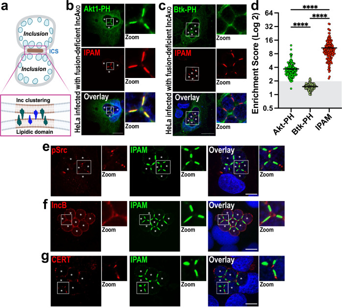
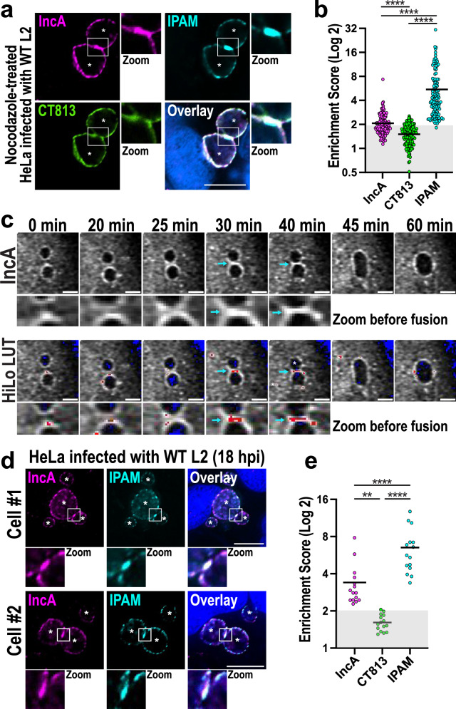
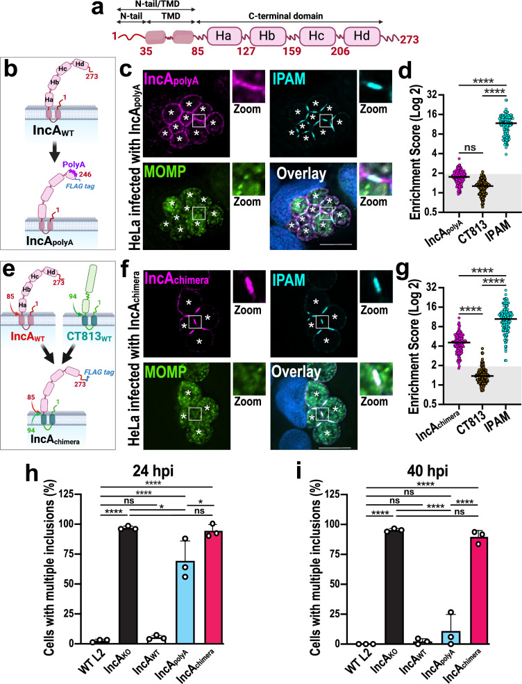
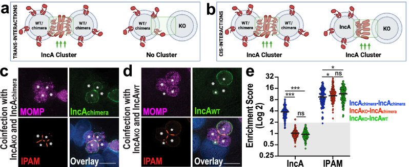
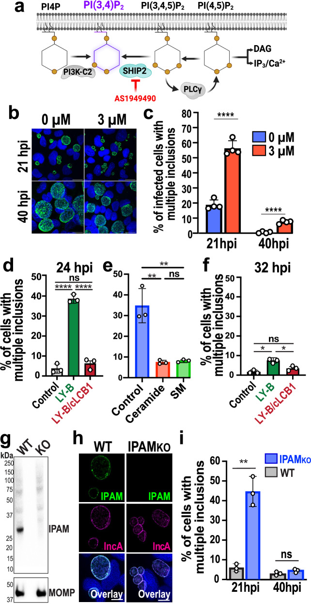
The intracellular bacterial pathogen Chlamydia trachomatis replicates within a membrane-bound compartment called the inclusion. Upon infection with several chlamydiae, each bacterium creates its own inclusion, resulting in multiple inclusions within each host cell. Ultimately, these inclusions fuse together in a process that requires the chlamydial protein IncA. Here, we show that inclusions form unique contact sites (inclusion contact sites, ICSs) prior to fusion, that serve as fusogenic platforms in which specific lipids and chlamydial proteins concentrate. Fusion depends on IncA clustering within ICSs and is regulated by PI(3,4)P2 and sphingolipids. As IncA concentrates within ICSs, its C-terminus likely interacts in trans with IncA on the apposing membrane, securing a high concentration of IncA at fusion sites. This regulatory mechanism contrasts with eukaryotic or viral fusion systems that are either composed of multiple proteins or use a change in pH to initiate membrane fusion. Thus, our study demonstrates that Chlamydia-mediated membrane fusion is primarily regulated by specific structural domains in IncA and its local organization on the inclusion membrane, which is affected by the host cell lipid composition.


Conflict of interest statement

The authors declare no competing interests.

Figures

Fig. 1
Fig. 1. Inclusions form unique contact sites (ICSs).
a Schematic depicts ICSs as points of contact between inclusion membranes in which specific lipids (brown) and proteins (green and blue) are clustered (see insert). Created in BioRender. Paumet, F. (2023) BioRender.com/s94s603. bd HeLa cells were infected with IncAKO Ctr (MOI 3). At 2 hpi, cells were transfected with plasmids expressing Akt-PH-GFP (b, green) or Btk-PH-GFP (c, green). Cells were then fixed at 24 hpi and labeled with anti-IPAM antibody (red). DNA was stained with Hoechst (blue). Enlarged ICSs (white box) are shown in the Zoom panel. Scale bar = 20 μm. Images are representative of three independent experiments. d ESs were calculated and plotted for each probe and IPAM as a control. Each dot = individual ES, black horizontal bar = mean. Gray box denotes ESs <2. n = 150 total scores per probe from three independent experiments. ns = not significant, **** denotes p-value < 0.0001 (one-way ANOVA). eg IncAKO Ctr infected cells were fixed at 24 hpi and labeled with anti-IPAM (green) and anti-pSrc (e), anti-IncB (f), or anti-CERT (g) antibodies (red). DNA was stained with Hoechst (blue). Enlarged ICSs (white box) are shown in the Zoom panel. Scale bar = 10 μm. Asterisks mark the inclusions. Images are representative of three independent experiments.
Fig. 2
Fig. 2. The bacterial fusion protein IncA clusters in ICSs.
a HeLa cells were infected with WT Ctr (MOI 3) for 5 h to allow the infection to proceed before being treated with 20 μM nocodazole. Cells were then fixed at 21 hpi and labeled with anti-IncA (magenta), anti-IPAM (cyan), and anti-CT813 (green) antibodies. DNA was labeled with Hoechst (blue). Enlarged ICSs (white box) are shown in the Zoom panel. Asterisks denote inclusions. Scale bar = 10 µm. Images are representative of three independent experiments. b ESs were calculated for each Inc. Each dot = individual ES, black horizontal bar = mean. Gray box denotes ESs <2. N = 130 total scores per Inc from three independent experiments. ns = not significant, **** denotes p-value < 0.0001 (matched-paired, one-way ANOVA). c Anti-FLAG-FrankenbodymScarlet3 HeLa cells were infected with IncAKO Ctr complemented with IncAWT-FLAG (MOI 3). IncAWT-FLAG expression was induced at 5 hpi with 20 ng/mL anhydrotetracycline. z-stacks of infected cells were acquired every 5 min as described in the Methods. Individual frames are shown beginning at 17 hpi (0 min). IncA images show the enrichment of the anti-FLAG FrankenbodymScarlet3 signal at the ICS prior to the fusion of the two inclusions (zoom). The HiLo look-up table (HiLo LUT) was then applied to the IncA images to highlight the grayscale differences (low signal = blue; high signal = red). Scale bar = 5 µm. Blue arrows denote signal enrichment at the ICS. Images are representative of three independent experiments. d HeLa cells were infected with WT Ctr (MOI 3), fixed at 18 hpi, and labeled with anti-IPAM (cyan) and -IncA (magenta) antibodies. DNA was labeled with Hoechst (blue). Enlarged ICSs (white box) are shown in the Zoom panel. Asterisks denote inclusions; Scale bar = 10 μm. Images are representative of three independent experiments. e HeLa cells were infected, fixed, and labeled as in d. ESs for each Inc was measured. N = 15 total scores per Inc from three independent experiments. Gray box denotes ESs <2. ns = not significant, **** denotes p-value < 0.0001 (matched-paired, one-way ANOVA).
Fig. 3
Fig. 3. The C-terminal domain of IncA controls IncA clustering in ICSs and regulates homotypic fusion.
a Schematic of full-length IncA, displaying an N-terminal tail (N-tail) and transmembrane domain (TMD), as well as four C-terminal helices (Ha-d). The N-tail and the C-terminal domain face the host cytoplasm, while the TMD anchors IncA in the inclusion membrane. Created in BioRender. Paumet, F. (2023) BioRender.com/z33h938. b Schematic of IncApolyA-FLAG in which Hd residues were mutated to alanines to disrupt critical intramolecular contacts to create the IncApolyA-FLAG complemented mutant strain. Created in BioRender. Paumet, F. (2023) BioRender.com/u70l441. c HeLa cells were infected with IncApolyA-FLAG Ctr (MOI 5) for 24 h in the presence of anhydrotetracycline. Cells were fixed and labeled with anti-FLAG (magenta, labels IncApolyA-FLAG), anti-IPAM (cyan), and anti-MOMP (green) antibodies. DNA was stained with Hoechst (blue). Asterisks denote the inclusions; Scale bar = 10 μm. Images are representative of three independent experiments. d ESs for cells infected with IncApolyA-FLAG Ctr at 24 hpi. N = 150 total scores per Inc from three independent experiments. Gray box denotes ESs <2. ns = not significant, **** denotes p-value < 0.0001 (one-way ANOVA). e Schematic of IncAchimera-FLAG in which IncA N-tail and TMD were swapped for those of CT813, to create the IncAchimera-FLAG complemented mutant strain. Created in BioRender. Paumet, F. (2024) BioRender.com/f62h052. f Cells were infected with IncAchimera-FLAG Ctr (MOI 5) for 24 h, fixed, and labeled as in c. Asterisks denote inclusions; Scale bar = 10 μm. Images are representative of three independent experiments. g ESs were calculated as in d. **** denotes p-value < 0.0001 (one-way ANOVA). h, i Homotypic fusion was quantified for the indicated strains in infected cells (MOI 7) at 24 hpi (h) and 40 hpi (i). Graphs depict the average number of cells containing multiple inclusions +/- SD from three independent experiments. ns = not significant, * denotes p-value < 0.05, **** denotes p-value < 0.0001 (one-way ANOVA).
Fig. 4
Fig. 4. IncA clustering in ICSs requires the presence of IncA on both membranes.
a If IncA interacts in trans, then clustering will only be observed when IncA is present on both inclusion membranes. WT = wild-type. KO = IncA knockout. Created in BioRender. Paumet, F. (2024) BioRender.com/w90v943. b If IncA interacts in cis, then IncA clustering will be observed when IncA is on one or both inclusion membranes. Created in BioRender. Paumet, F. (2024) BioRender.com/q49y738. c, d HeLa cells were co-infected with IncAKO Ctr (MOI 2.5) and IncAchimera-FLAG Ctr (MOI 2.5) (c), or with IncAKO Ctr (MOI 2.5) and WT Ctr (MOI 2.5) (d) in the presence of anhydrotetracycline. Cells were fixed at 24 hpi and labeled with anti-MOMP (magenta), which labels Ctr, anti-IncA (green), and anti-IPAM (red) antibodies. DNA was stained with Hoechst (blue). For c, d: Asterisks denote inclusions; Scale bars = 10 μm. Images are representative of three independent experiments. e ES quantification for the co-infections conducted in c and d. Graph depicts the average (black line) and individual ESs (dots). n = 150 total scores per Inc from three independent experiments. Gray box denotes ESs <2. ns = not significant, * denotes p-value < 0.05, *** denotes p-value < 0.001 (two-way ANOVA).
Fig. 5
Fig. 5. The lipid and protein composition of the ICSs regulate homotypic fusion.
a Diagram of PI(3,4)P2 biosynthesis. The inhibitor AS1949490 used to block SHIP2 is indicated in red. Created in BioRender. Paumet, F. (2024) BioRender.com/p53j299. b HeLa cells were infected with WT Ctr (MOI 3) in the presence of 0 µM or 3 µM AS1949490. Cells were fixed at 21 hpi and labeled with anti-MOMP antibody (green) to visualize inclusions. Hoechst was used to label DNA (blue). Scale bar = 20 µm. Images are representative of four independent experiments. c Average percentage of cells treated with 0 µM or 3 µM AS1949490 containing multiple inclusions +/- SD from four independent experiments. 100 cells were counted from multiple fields of view for each experiment and each condition. **** denotes p-value < 0.0001 (two-tailed t-test). d, f CHO (control), LY-B, and LY-B/cLCB1 cells were infected with WT Ctr (MOI 3) and fixed at 24 or 32 (hpi). The average percentage of cells containing multiple inclusions was measured in WT Ctr-infected cells at 24 hpi (d) or 32 hpi (f) +/- SD from three independent experiments. For each replicate, 100 cells for each condition were counted from multiple fields of view. ns = not significant, * denotes p-value < 0.05, ** denotes p-value < 0.01, and **** denotes p-value < 0.0001 (one-way ANOVA). e LY-B cells were infected with WT Ctr (MOI 3) for 2 h followed by the addition of media supplemented with dihydroceramide (ceramide) or sphingomyelin (SM). Cells were then fixed at 24 hpi, and fusion was quantified as in d. g Western blot for the detection of IPAM in HeLa cell lysates infected with WT or IPAMKO Ctr (MOI 1, 24 hpi) shows successful knock out of IPAM (predicted molecular weight 29.4kD) in IPAMKO Ctr. MOMP staining is used as the loading control. Representative of two independent experiments. h HeLa cells were infected with WT or IPAMKO Ctr (MOI 3) and fixed 21 hpi. Cells were labeled with anti-IPAM (green) and anti-IncA (magenta) antibodies. DNA was stained with Hoechst (blue). Scale bar = 10 μm. Images are representative of two independent experiments. i Fusion was quantified as in d at 21 or 40 hpi.
Fig. 6
Fig. 6. Regulatory model of IncA-mediated membrane fusion.
❶ IncA (pink) is homogeneously expressed on the inclusion membrane where it is folded as a monomer to prevent cis self-assembly. IPAM (green) is distributed throughout the inclusion membrane. ❷ Lipids (blue), in particular sphingolipids and PI(3,4)P2, trigger IncA clustering within ICSs. PI(3,4)P2 also controls IPAM recruitment into the ICSs. ❸ As IncA concentrates in the ICS, it then oligomerizes in trans, further enhancing its clustering. ❹ At its optimal local concentration, IncA drives the homotypic fusion of inclusions. Ct = Chlamydia trachomatis bacteria. Created in BioRender. Paumet, F. (2023) BioRender.com/c27a297.

References

    1. Mellman, I. & Warren, G. The road taken: past and future foundations of membrane traffic. Cell100, 99–112 (2000). - PubMed
    1. Blumenthal, R., Clague, M. J., Durell, S. R. & Epand, R. M. Membrane fusion. Chem. Rev.103, 53–69 (2003). - PubMed
    1. Fischer, A. & Rudel, T. Safe haven under constant attack-the Chlamydia-containing vacuole. Cell Microbiol.20, e12940 (2018). - PubMed
    1. Hackstadt, T., Scidmore-Carlson, M. A., Shaw, E. I. & Fischer, E. R. The Chlamydia trachomatis IncA protein is required for homotypic vesicle fusion. Cell. Microbiol.1, 119–130 (1999). - PubMed
    1. Weber, M. et al. A functional core of IncA is required for Chlamydia trachomatis inclusion fusion. J. Bacteriol.198, 1347–1355 (2016). - PMC - PubMed

Publication types