

Diagnosis of canine B-cell chronic lymphoid leukemia with a CD21 negative phenotype using the LT21 clone CD21 antibody in flow cytometry: a case report
- PMID: 39462364
- PMCID: PMC11515120
- DOI: 10.1186/s12917-024-04335-x
Diagnosis of canine B-cell chronic lymphoid leukemia with a CD21 negative phenotype using the LT21 clone CD21 antibody in flow cytometry: a case report
Abstract
Background: Chronic lymphoid leukemia (CLL) is a hematological disorder characterized by the clonal expansion of small mature lymphocytes that accumulate in the blood and bone marrow. CLL can arise from B-, T-, or natural killer cell clones. The cytological evaluation of blood smears is often the simplest and least invasive method for diagnosing lymphoid leukemia. Immunophenotyping is used to further subclassify the type of lymphoid leukemia.
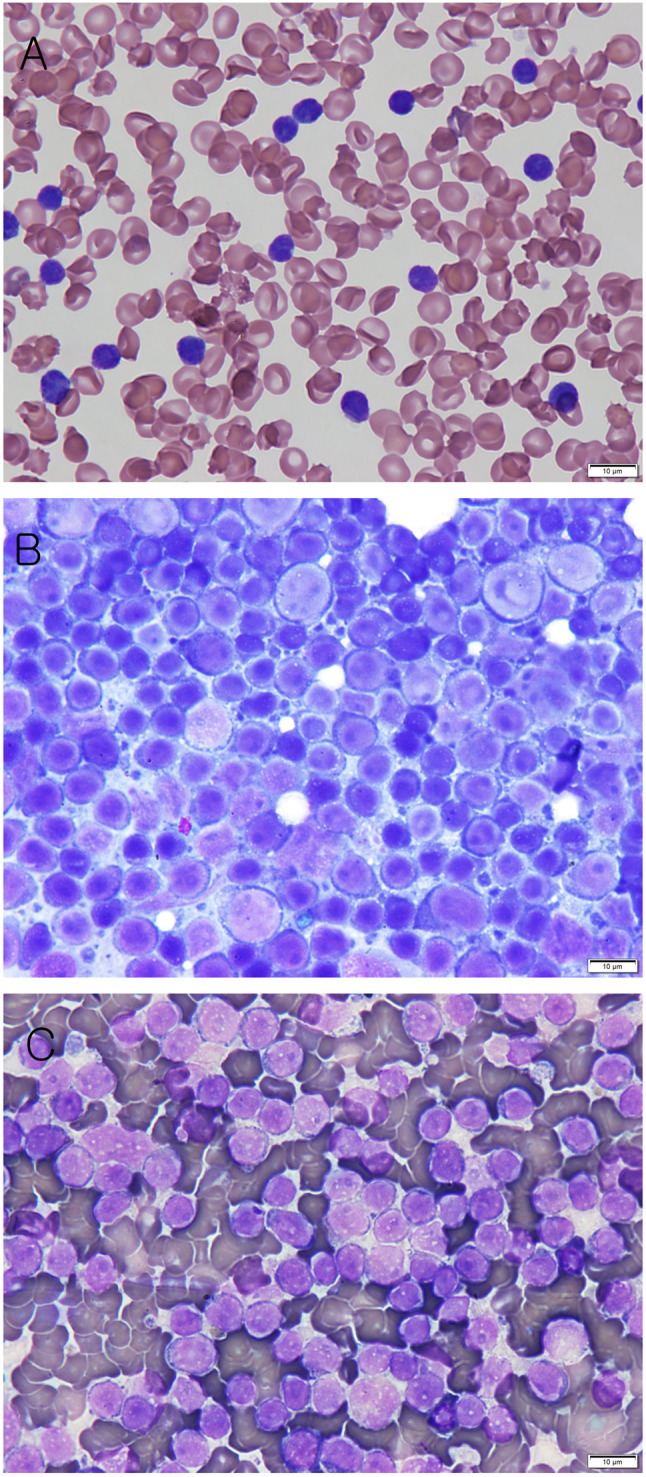
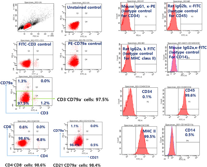
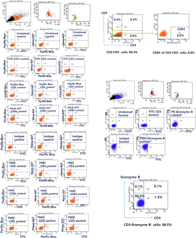
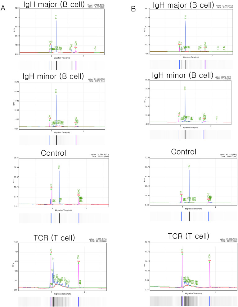
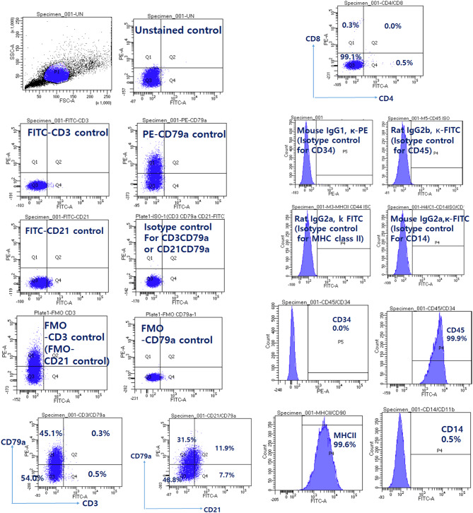
Case presentation: A 15-year-old, 4.4-kg spayed female Shih Tzu was presented to the veterinary medical teaching hospital of Kangwon National University. Despite having a normal appetite and activity level, cervical and inguinal lymph node enlargement was noted on physical examination. Complete blood count revealed severe leukocytosis, severe lymphocytosis, and monocytosis. Splenomegaly, hepatomegaly, and lymph node enlargement were detected on radiographic and ultrasonographic examination. Immunophenotyping was performed using peripheral blood mononuclear cells (PBMCs). The majority of lymphocytes exhibited the following profiles: CD3-CD79a- (97.5%), CD4-CD8- (98.6%), CD21-CD79a- (98.4%), CD34- (0.1%), CD45+ (99.6%), major histocompatibility complex class II+ (99.5%), and CD14- (0.5%). Based on the immunophenotyping results, possible differentials considered included the following: the majority of lymphocytes may be natural killer (NK) cell clones, plasma cell clones, or show aberrant expression or loss of CD21 marker due to the neoplastic nature of the cells. Further flow cytometry was performed using antibodies against CD3, CD5, CD94, and granzyme B. The combined results indicated that the predominant lymphocyte subset in the PBMCs was CD3-CD5-CD21-CD94-granzyme B-. To confirm monoclonality and exclude the aberrant loss of CD markers, a polymerase chain reaction for antigen receptor rearrangement (PARR) assay was conducted. The PARR assay, using DNA from blood and lymph node samples, showed B-cell monoclonality. Immunocytochemistry using PBMCs showed that the plasma cell marker Multiple Myeloma Oncogene 1 (MUM1) was not expressed. Therefore, the diagnosis was confirmed to be B-cell CLL.
Conclusion: Immunophenotyping can help subclassify the type of lymphoid leukemia; however, as tumor cells can show aberrant expression or loss of the CD21 marker, combining immunophenotyping with the PARR assay could yield a more accurate diagnosis.
Keywords: B-cell chronic lymphoid leukemia; CD21; Dog; Immunophenotyping; Lymphoproliferative diseases.
© 2024. The Author(s).
Conflict of interest statement
The authors declare no competing interests.
Figures
References
-
- Comazzi S, Gelain ME, Martini V, Riondato F, Miniscalco B, Marconato L, Stefanello D, Mortarino M. Immunophenotype predicts survival time in dogs with chronic lymphocytic leukemia. J Vet Intern Med. 2011;25:100–6. - PubMed
-
- Zabolotzky SM, Walker DB. Chapter 26. Peripheral blood smears. In: Valenciano AC, Cowell RL, editors. Cowell and Thyler’s Diagnostic Cytology and Hematology of the dog and cat. 4th ed. Beijing, China: Elsevier; 2014. pp. 483–6.
-
- Presley RH, Mackin A, Vernau W. Lymphoid leukemia in dogs. Compendium Continuing Educ Practicing Veterinarian. 2006;28:831–49.
-
- Vernau W, Moore PF. An Immunophenotypic Study of Canine Leukemias and Preliminary Assessment of Clonality by polymerase chain reaction. Vet Immunol Immunopathol. 1999;69:145–64. - PubMed
Publication types
MeSH terms
Substances
LinkOut - more resources
Full Text Sources
Research Materials
Miscellaneous
