. 2024 Oct 26;22(1):662.
doi: 10.1186/s12951-024-02830-9.

S-RBD-modified and miR-486-5p-engineered exosomes derived from mesenchymal stem cells suppress ferroptosis and alleviate radiation-induced lung injury and long-term pulmonary fibrosis

Affiliations

S-RBD-modified and miR-486-5p-engineered exosomes derived from mesenchymal stem cells suppress ferroptosis and alleviate radiation-induced lung injury and long-term pulmonary fibrosis

Wei-Yuan Zhang et al. J Nanobiotechnology. .

Abstract

Background: Radiation-induced lung injury (RILI) is associated with alveolar epithelial cell death and secondary fibrosis in injured lung. Mesenchymal stem cell (MSC)-derived exosomes have regenerative effect against lung injury and the potential to intervene of RILI. However, their intervention efficacy is limited because they lack lung targeting characters and do not carry sufficient specific effectors. SARS-CoV-2 spike glycoprotein (SARS-CoV-2-S-RBD) binds angiotensin-converting enzyme 2 (ACE2) receptor and mediates interaction with host cells. MiR-486-5p is a multifunctional miRNA with angiogenic and antifibrotic potential and acts as an effector in MSC-derived exosomes. Ferroptosis is a form of cell death associated with radiation injury, its roles and mechanisms in RILI remain unclear. In this study, we developed an engineered MSC-derived exosomes with SARS-CoV-2-S-RBD- and miR-486-5p- modification and investigated their intervention effects on RIPF and action mechanisms via suppression of epithelial cell ferroptosis.

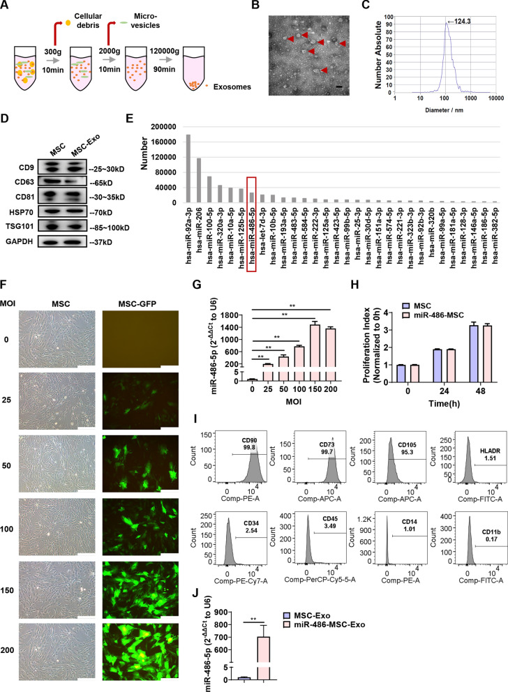
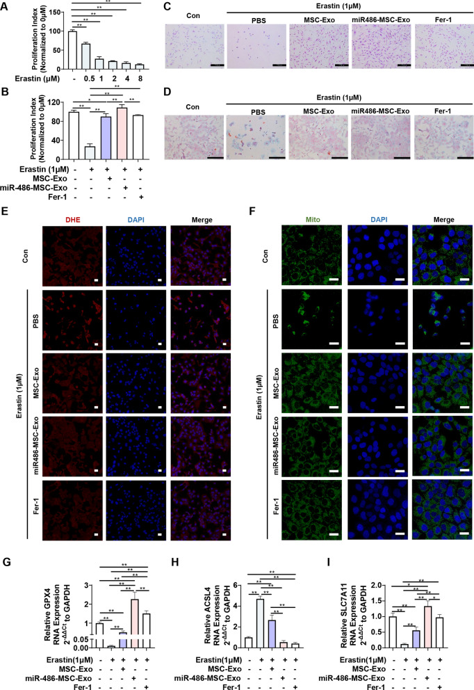
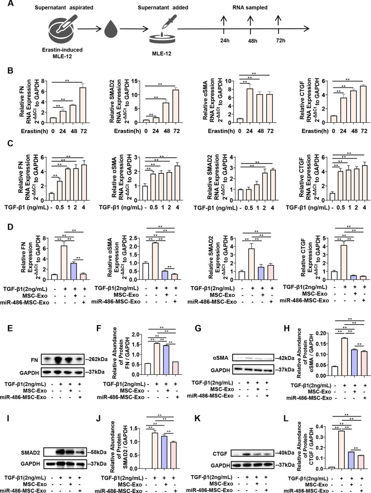
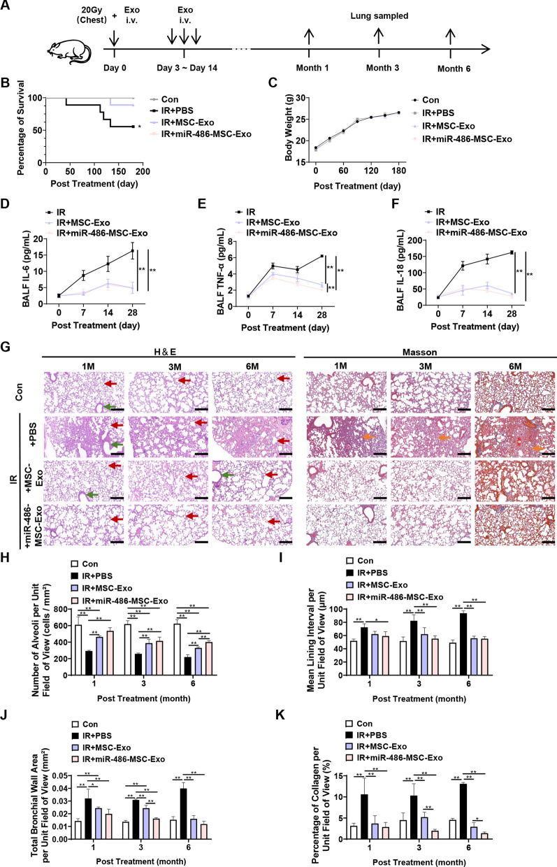
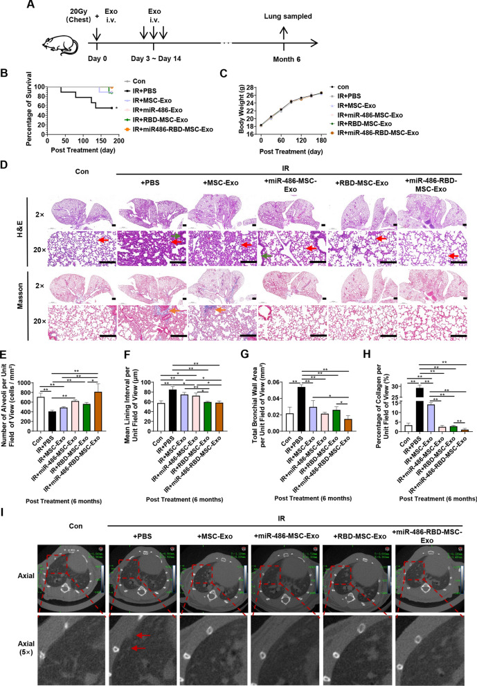
Results: Adenovirus-mediated gene modification led to miR-486-5p overexpression in human umbilical cord MSC exosomes (p < 0.05), thereby constructing miR-486-5p engineered MSC exosomes (miR-486-MSC-Exo). MiR-486-MSC-Exo promoted the proliferation and migration of irradiated mouse lung epithelial (MLE-12) cells in vitro and inhibited RILI in vivo (all p < 0.05). MiR-486-MSC-Exo suppressed ferroptosis in MLE-12 cells, and an in vitro assay revealed that the expression of fibrosis-related genes is up-regulated following ferroptosis (both p < 0.05). MiR-486-MSC-Exo reversed the up-regulated expression of fibrosis-related genes induced by TGF-β1 in vitro and improved pathological fibrosis in RIPF mice in vivo (all p < 0.05). SARS-CoV-2-S-RBD-modified and miR-486-5p-engineered MSC exosomes (miR-486-RBD-MSC-Exo) were also constructed, and the distribution of DiR dye-labeled miR-486-RBD-MSC-Exo in hACE2CKI/CKI Sftpc-Cre+ mice demonstrated long-term retention in the lung (p < 0.05). MiR-486-RBD-MSC-Exo significantly improved the survival rate and pathological changes in hACE2CKI/CKI Sftpc-Cre+ RIPF mice (all p < 0.05). Furthermore, miR-486-MSC-Exo exerted anti-fibrotic effects via targeted SMAD2 inhibition and Akt phosphorylation activation (p < 0.05).

Conclusions: Engineered MSC exosomes with SARS-CoV-2-S-RBD- and miR-486-5p-modification were developed. MiR-486-RBD-MSC-Exo suppressed ferroptosis and fibrosis of MLE-12 cells in vitro, and alleviated RILI and long-term RIPF in ACE2 humanized mice in vivo. MiR-486-MSC-Exo exerted anti-fibrotic effects via SMAD2 inhibition and Akt activation. This study provides a potential approach for RIPF intervention.

Keywords: Engineered exosomes; Ferroptosis; Mesenchymal stem cells; MiR-486-5p; Pulmonary fibrosis; Radiation-induced pulmonary injury; SARS-CoV-2-S-RBD.


Conflict of interest statement

The authors declare no competing interests.

Figures

Fig. 1
Fig. 1
The construction and biological characteristics of miR-486-5p-engineered MSC exosomes. A Schematic representation of exosome extraction using ultracentrifugation. B A representative transmission electron microscopy image of exosomes, with arrows indicating typical exosomes. Scale bar = 200 nm. C Nanoparticle tracking analysis of exosomes. D Western blot analysis of MSCs (MSC) and MSC-derived exosomes (MSC-Exo). The protein expression of HSP70, CD81, CD63 and TSG101 were detected and GAPDH was used as an internal control. E Top 30 small RNAs identified using exosomal sequencing. F Green fluorescence infected with gradient multiplicity of infection (MOI) of Ad-GFP-miR-486-5p adenovirus. Scale bar = 200 μm. G Expression of miR-486-5p in miR-486-5p-engineered MSC (miR-486-MSC) determined using qPCR. Data are expressed as the mean ± SD. *p < 0.05, **p < 0.01. H Comparison of the proliferation activity of miR-486-MSC and MSC. I Identification of CD90, CD73, CD105, CD45, CD34, and CD14 expression on the surface of miR-486-MSC using flow cytometry. J Expression of miR-486-5p in miR-486-5p-engineered MSC-exosomes (miR-486-MSC-Exo) compared with MSC-exosomes (MSC-Exo). Data are expressed as the mean ± SD. *p < 0.05, **p < 0.01
Fig. 2
Fig. 2
MiR-486-5p-engineered MSC exosomes increased proliferation and migration of MLE-12 cells after irradiation. A Proliferation of MLE-12 cells treated with MSC-Exo or miR-486-MSC-Exo at 0, 24, 48, 72, and 96 h using CCK-8 assay. 1 × PBS was used as the control. Data are expressed as the mean ± SD. *p < 0.05, **p < 0.01. B Proliferation of MLE-12 cells treated with 6, 8, 10, or 12 Gy γ-rays irradiation at 0, 24, 48, 72 and 96 h using CCK-8 assay. 0 h cells were used as control. Data are expressed as the mean ± SD. *p < 0.05, **p < 0.01. C Proliferation of MLE-12 cells treated with MSC-Exo or miR-486-MSC-Exo at 0, 24, 48, 72, and 96 h after irradiation with 10 Gy γ-rays using CCK-8 assay. The irradiated cells served as a control. Data are expressed as the mean ± SD. *p < 0.05, **p < 0.01. D Proliferation of MLE-12 cells transduced with miR-NC or miR-486-5p at 0, 24, 48, 72, and 96 h after irradiation with 10 Gy γ-rays using CCK-8 assay. The irradiated cells served as a control. Data are expressed as the mean ± SD. *p < 0.05, **p < 0.01. E Morphology of MLE-12 cells treated with MSC-Exo or miR-486-MSC-Exo at 48 h after irradiation with 10 Gy γ-rays using Giemsa staining, bar = 200 μm. F-G Scratch assay to assess the migration of MLE-12 cells treated with MSC-Exo or miR-486-MSC-Exo. Representative images and quantitative analysis rate at 0, 6, 12, and 24 h were shown. Scale bar = 200 μm. Data are expressed as the mean ± SD. *p < 0.05, **p < 0.01
Fig. 3
Fig. 3
MiR-486-5p engineered MSC exosomes alleviated erastin-induced ferroptosis in MLE-12 cells. A Cell viability of MLE-12 cells treated with increasing doses of erastin (from 0.5 to 8 μM) at 24 h using CCK-8 assay. Data are expressed as the mean ± SD. *p < 0.05, **p < 0.01. B Cell viability (24 h) of MLE-12 cells treated with MSC-Exo or miR-486-MSC-Exo after adding erastin (1 μM) using CCK-8 assay. Fer-1 was used as a positive control. Data are expressed as the mean ± SD. *p < 0.05, **p < 0.01. C Morphological changes (24 h) and D divalent iron accumulation (24 h) of MLE-12 cells treated with MSC-Exo or miR-486-MSC-Exo after adding erastin (1 μM) using giemsa staining and divalent iron staining. Fer-1 was used as a positive control. E Dihydroethidine (DHE) (24 h) and F mitochondrial fluorescence probe (mito-tracker) labeling (24 h) of MLE-12 cells treated with MSC-Exo or miR-486-MSC-Exo after adding erastin (1 μM). Fer-1 was used as a positive control. Scale bar = 10 μm G-I mRNA expression (24 h) of GPX4, ACSL4, and SLC7A11 in MLE-12 cells treated with MSC-Exo or miR-486-MSC-Exo after adding erastin (1 μM) using qPCR. Fer-1 was used as a positive control. Data are expressed as the mean ± SD. *p < 0.05, **p < 0.01
Fig. 4
Fig. 4
MiR-486-5p-engineered MSC exosomes reduce pulmonary fibrosis in vitro. A-B mRNA expression of FN, αSMA, SMAD2, and CTGF in MLE-12 cells treated with erastin-induced supernatant at 24 h, 48 h, and 72 h via qPCR. Data are expressed as the mean ± SD. *p < 0.05, **p < 0.01. C mRNA expression of FN, αSMA, SMAD2, and CTGF in MLE-12 cells treated with gradient concentrations (0, 1, 2, 4 ng/mL) of TGF-β1. Data are expressed as the mean ± SD. *p < 0.05, **p < 0.01. D mRNA expression of FN, αSMA, SMAD2, and CTGF in MLE-12 cells treated with MSC-Exo or miR-486-MSC-Exo after adding TGF-β1 (2 ng/mL). Data are expressed as the mean ± SD. *p < 0.05, **p < 0.01. E-L Protein expression and quantitative analysis of FN, αSMA, SMAD2, and CTGF in MLE-12 cells with MSC-Exo or miR-486-MSC-Exo after adding TGF-β1 (2 ng/mL), with GAPDH as the internal reference. Data are expressed as the mean ± SD. *p < 0.05, **p < 0.01
Fig. 5
Fig. 5
MiR-486-5p engineered MSC exosomes reduce pulmonary fibrosis in vivo. A Schematic representation. B Survival curves and C body weight changes of RIPF mice treated with MSC-Exo or miR-486-MSC-Exo, with nine mice in each group. 1 × PBS was used as negative control. D-F Expression of IL-6, TNF-α, and IL-18 in the bronchoalveolar lavage fluid (BALF) of RIPF model mice treated with MSC-Exo or miR-486-MSC-Exo via ELISA. Data are expressed as the mean ± SD. *p < 0.05, **p < 0.01. G Representative images of H&E and Masson staining at 1 M, 3 M, or 6 M in RIPF mice treated with MSC-Exo or miR-486-MSC-Exo, with four mice in each group. 1 × PBS was used as negative control. The red, green, and orange arrows indicate alveoli, bronchial walls, and collagen, respectively. Scale bar = 200 μm. H–K Statistical analysis of H&E and masson, including number of alveoli per unit field of view, mean lining interval per unit field of view, total bronchial wall area per unit field of view, and percentage of collagen per unit field of view. Data are expressed as the mean ± SD. *p < 0.05, **p < 0.01
Fig. 6
Fig. 6
Exosomes can achieve targeted lung delivery in vivo via RBD-hACE2. A Structure diagram of RBD-miR-486-5p engineered exosomes (miR-486-RBD-MSC-Exo). B Western blot analysis of RBD protein expression in RBD-MSC and RBD-MSC-Exo, with MSC as a comparison. GAPDH was used as the internal reference. C Construction diagram of hACE2CKI/CKI -SftpcCre+ mice. D Genotype identification of hACE2CKI/CKI -SftpcCre+ mice. E Western blot analysis of hACE2 expression in hACE2CKI/CKI -SftpcCre mice and hACE2CKI/CKI -SftpcCre+ mice with tamoxifen induction. β-actin was used as the internal reference. F Early organ distribution of miR-486-RBD-MSC-Exo bound to DiR after intravenous administration in hACE2CKI/CKI -SftpcCre+ mice. Scramble-miR-486-5p engineered exosomes (miR-486-Scamble-MSC-Exo) was used as control. G-I Quantitative analysis of fluorescence distribution from E. Data are expressed as the mean ± SD. *p < 0.05, **p < 0.01
Fig. 7
Fig. 7
The targeted binding of RBD-hACE2 in miR-486-5p-engineered MSC exosomes enhance treatment efficacy for pulmonary fibrosis. A Schematic representation. B Survival curves of hACE2CKI/CKI -SftpcCre+ mice treated with MSC-Exo, miR-486-MSC-Exo, RBD-MSC-Exo and miR-486-RBD-MSC-Exo after irradiation, with nine mice in each group. 1 × PBS was used as negative control. C Body weight changes of RIPF mice treated with MSC-Exo or miR-486-MSC-Exo, with nine mice in each group. 1 × PBS was used as negative control. D Representative images of H&E and Masson staining at 6 M in RIPF hACE2CKI/CKI -SftpcCre+ mice treated with MSC-Exo, miR-486-MSC-Exo, RBD-MSC-Exo and miR-486-RBD-MSC-Exo, with four mice in each group. 1 × PBS was used as negative control. Scale bar = 200 μm. E–H Statistical analysis of H&E and masson, including number of alveoli per unit field of view, mean lining interval per unit field of view, total bronchial wall area per unit field of view, and percentage of collagen per unit field of view. The red, green, and orange arrows indicate alveoli, bronchial walls, and collagen, respectively. Data are expressed as the mean ± SD. *p < 0.05, **p < 0.01. I Representative small animal CT images in RIPF hACE2CKI/CKI -SftpcCre+ mice treated with MSC-Exo, miR-486-MSC-Exo, RBD-MSC-Exo and miR-486-RBD-MSC-Exo. The red arrow indicates areas of fibrosis. 1 × PBS was used as negative control
Fig. 8
Fig. 8
MiR-486-5p-engineered MSC exosomes alleviates pulmonary fibrosis via miR-486-5p-SMAD2-pAkt. A SMAD2 protein expression in lungs of RIPF hACE2CKI/CKI -SftpcCre+ mice at 6 M after irradiation via immunofluorescence, with four mice in each group. Representative images were shown. Scale bar = 200 μm. B Fluorescent quantitation of SMAD2 protein expression (red) in RIPF mice treated with different exosomes. Data are expressed as the mean ± SD. *p < 0.05, **p < 0.01. C mRNA and D-E protein expression of SMAD2 in MLE-12 cells treated with miR-486-5p mimic (miR-486-5p) after adding TGF-β1 (2 ng/mL). MiR-NC was used as control. Data are expressed as the mean ± SD. *p < 0.05, **p < 0.01. F MiR-486-5p target site in the SMAD2 3' UTR and the sequence alignment of miR-486-5p and the SMAD2 3' UTR. Mutated bases in the psiCHECK-2 construct are bold. G Luciferase activity after co-transfection of miR-486-5p and SMAD2 into HEK293 cells. MiR-NC was used as a control. Renilla luciferase activity was normalized to firefly luciferase activity. Data are expressed as the mean ± SD. *p < 0.05, **p < 0.01. H-J Western blot analysis of Akt phosphorylation in MLE-12 cells treated with MSC-Exo or miR-486-MSC-Exo. Insulin was used as an inducer of Akt phosphorylation. LY294002 was used as an inhibitor of Akt phosphorylation. Data are expressed as the mean ± SD. *p < 0.05, **p < 0.01

References

    1. Hanania AN, Mainwaring W, Ghebre YT, Hanania NA, Ludwig M. Radiation-induced lung injury: assessment and management. Chest. 2019;156:150–62. - PMC - PubMed
    1. Yan Y, Fu J, Kowalchuk RO, Wright CM, Zhang R, Li X, Xu Y. Exploration of radiation-induced lung injury, from mechanism to treatment: a narrative review. Transl Lung Cancer Res. 2022;11:307–22. - PMC - PubMed
    1. Kim N, Cho SG. Clinical applications of mesenchymal stem cells. Korean J Intern Med. 2013;28:387–402. - PMC - PubMed
    1. Zanoni M, Cortesi M, Zamagni A, Tesei A. The role of mesenchymal stem cells in radiation-induced lung fibrosis. Int J Mol Sci. 2019;20(16):3876. - PMC - PubMed
    1. Xu R, Feng Z, Wang FS. Mesenchymal stem cell treatment for COVID-19. EBioMedicine. 2022;77: 103920. - PMC - PubMed

LinkOut - more resources