. 2024 Oct 26;12(1):31.
doi: 10.1186/s40170-024-00361-3.

GCN2-SLC7A11 axis coordinates autophagy, cell cycle and apoptosis and regulates cell growth in retinoblastoma upon arginine deprivation

Affiliations

GCN2-SLC7A11 axis coordinates autophagy, cell cycle and apoptosis and regulates cell growth in retinoblastoma upon arginine deprivation

Dan Wang et al. Cancer Metab. .

Abstract

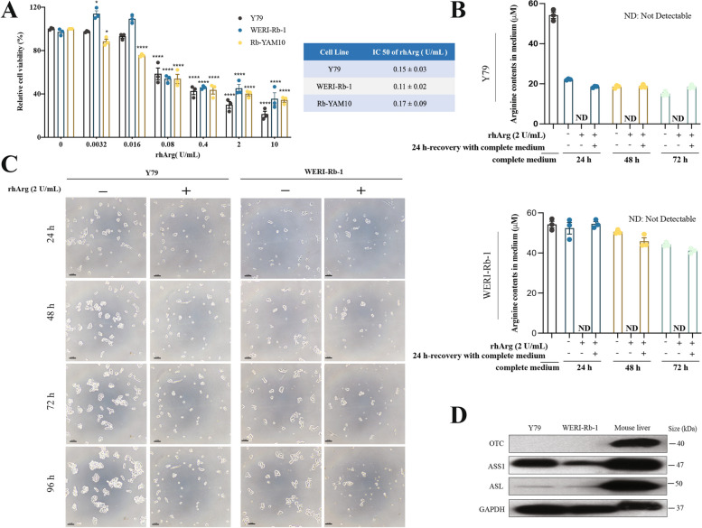
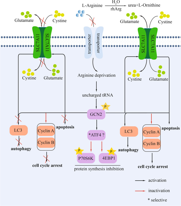
Background: Arginine deprivation was previously shown to inhibit retinoblastoma cell proliferation and induce cell death in vitro. However, the mechanisms by which retinoblastoma cells respond to arginine deprivation remain to be elucidated.

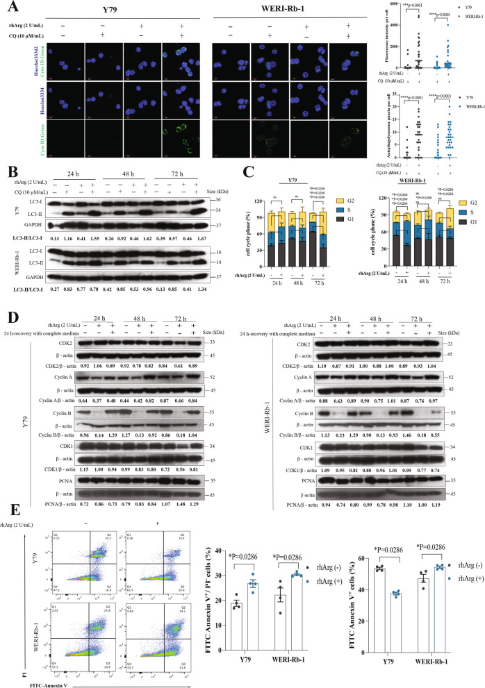
Methods: The human-derived retinoblastoma cell lines Y79 and WERI-Rb-1 were subjected to arginine depletion, and the effects on inhibiting cell growth and survival were evaluated. This study investigated potential mechanisms, including autophagy, cell cycle arrest and apoptosis. Moreover, the roles of the general control nonderepressible 2 (GCN2) and mechanistic target of rapamycin complex 1 (mTORC1) signaling pathways in these processes were examined.

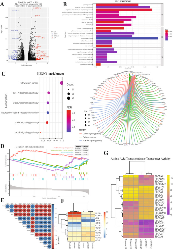
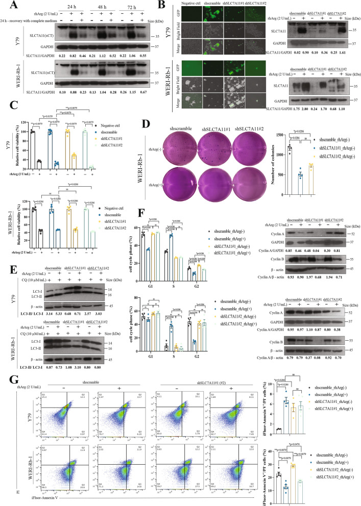
Results: We demonstrated that arginine deprivation effectively inhibited the growth of retinoblastoma cells in vitro. This treatment caused an increase in the autophagic response. Additionally, prolonged arginine deprivation induced G2 cell cycle arrest and was accompanied by an increase in early apoptotic cells. Importantly, arginine depletion also induced the activation of GCN2 and the inhibition of mTOR signaling. We also discovered that the activation of SLC7A11 was regulated by GCN2 upon arginine deprivation. Knockdown of SLC7A11 rendered retinoblastoma cells partially resistant to arginine deprivation. Furthermore, we found that knockdown of GCN2 led to a decrease in the autophagic response in WERI-Rb-1 cells and arrested more cells in S phase, which was accompanied by fewer apoptotic cells. Moreover, knockdown of GCN2 induced the constant expression of ATF4 and the phosphorylation of 70S6K and 4E-BP1 regardless of arginine deprivation.

Conclusions: Collectively, our findings suggest that the GCN2‒SLC7A11 axis regulates cell growth and survival upon arginine deprivation through coordinating autophagy, cell cycle arrest, and apoptosis in retinoblastoma cells. This work paves the way for the development of a novel treatment for retinoblastoma.

Keywords: Arginine deprivation; GCN2; Retinoblastoma; SCL7A11.


Conflict of interest statement

The authors declare no competing interests.

Figures

Fig. 1
Fig. 1
Arginine deprivation inhibited the growth of retinoblastoma cells in vitro. A Retinoblastoma cell lines (Y79, WERI-Rb-1, and Rb-YAM10) were treated with increasing concentrations of rhArg for 72 h. The data are presented as the means ± SEMs, n = 3. Differences were assessed by one-way ANOVA followed by Bonferroni’s multiple comparison test. p values are indicated as follows: *, p < 0.05; ****, p < 0.0001. B HPLC‒MS analysis of the arginine levels in the cell culture medium. n = 3. ND, not detected. C Growth pattern changes following arginine deprivation at the indicated time points. Scale bar, 100 μm. D Characterization of urea cycle enzymes in retinoblastoma. Mouse liver extract was used as the positive control
Fig. 2
Fig. 2
Arginine deprivation induced autophagy, cell cycle arrest and apoptosis in retinoblastoma, which were accompanied by GCN2 signaling activation and mTOR signaling inhibition. A Autophagy was quantified by Cyto-ID staining. Chloroquine (CQ), an autophagosome–lysosome fusion inhibitor; Hoechst 33342, stain for cell nuclei; Cyto-ID green, fluorescent dye that selectively labels accumulated autophagic vacuoles. The medians are indicated in the quantification plots. n = 20–27, which corresponds to the number of cells analyzed. B Immunoblot analyses showing time-dependent changes in autophagy activity (conversion of LC3-I to LC3-II) in Rb cells following arginine deprivation. C Flow cytometry analyses of cell cycle arrest. The bar chart shows the mean ± SEM of the percentage of cells in each cell cycle phase after arginine deprivation from 24– to 72 h (n = 4). ns = not significant. D Immunoblot analyses showing time-dependent changes in the expression of cell cycle-related proteins (CDK1, cyclin A, cyclin B and CDK2). E Flow cytometry analyses of early apoptotic cells. The bar charts show the mean ± SEM of the percentage of early apoptotic cells 72 h after arginine deprivation (n = 4)
Fig. 3
Fig. 3
Identification of candidate genes regulated by arginine deprivation in Y79 cells. A Volcano plot showing changes in gene expression due to arginine deprivation. B and C GO and KEGG enrichment analyses of differentially expressed genes (DEGs) (|fold change|> 1 and adjusted p values < 0.05) in Y79 cells. E Correlations between cell cycle-related genes. F Heatmap of cell cycle related–related genes. G Heatmap of the solute carrier (SLC) family
Fig. 4
Fig. 4
GCN2 regulated autophagy, cell cycle arrest and apoptosis in retinoblastoma upon arginine deprivation. A Immunoblot analyses revealed that GCN2 was induced upon arginine deprivation in both Y79 and WERI-Rb-1 cells. B Fluorescence microscopy image showing GFP signals in cells and immunoblot data showing the efficiency of GCN2 knockdown in Rb cells. Scale bar, 300 μm. C Y79 and WERI-Rb-1 cells were treated with rhArg for 72 h. The data are shown as the means ± SEMs, n = 4–5. Differences were assessed by the Mann‒Whitney test: ns = not significant. D Representative images and colony counts of GCN2-knockdown WERI-Rb-1 cells. E Immunoblot analyses showing the changes in autophagy activity (conversion of LC3-I to LC3-II) in Rb cells following arginine deprivation. F Flow cytometry analyses of cell cycle arrest. The bar chart shows the mean ± SEM of the percentage of cells in each cell cycle phase after 72 h of exposure of Rb cells to rhArg (n = 4). Immunoblot analyses showing the changes in cyclin A and cyclin B. G Flow cytometry analyses of apoptosis. The bar chart shows the mean ± SEM of the percentage of early apoptotic cells 72 h after arginine deprivation (n = 4)
Fig. 5
Fig. 5
SLC7A11 regulated autophagy, cell cycle arrest and apoptosis in retinoblastoma upon arginine restriction. A Immunoblot analyses revealed that SLC7A11 was induced upon arginine deprivation in both Y79 and WERI-Rb-1 cells. B Fluorescence microscopy image showing GFP signals in the cells and immunoblot data showing the efficiency of SLC7A11 knockdown in Rb cells. Scale bar, 300 μm. C Retinoblastoma cell lines (Y79, WERI-Rb-1) were treated with rhArg for 72 h. The data are shown as the means ± SEMs, n = 4–5. Differences were assessed by the Mann‒Whitney test: ns = not significant. D Representative images and colony counts of SLC7A11-knockdown WERI-Rb-1 cells. E Immunoblot analyses showing the changes in autophagy activity (conversion of LC3-I to LC3-II) in Rb cells after arginine deprivation. F Flow cytometric analyses of cell cycle arrest. The bar chart shows the mean ± SEM of the percentage of cells in the cell cycle phase after 72 h of exposure of Rb cells to rhArg (n = 4), and immunoblot analyses revealed changes in the expression of cell cycle-related proteins (cyclin A and cyclin B). G Flow cytometric analyses of cell apoptosis. The bar chart shows the mean ± SEM of the percentage of early apoptotic cells 72 h after arginine deprivation (n = 4)
Fig. 6
Fig. 6
Schematic summary. Mechanisms underlying the role of GCN2-SLC7A11 in regulating cell growth and survival through autophagy, cell cycle arrest and apoptosis in retinoblastoma cells upon arginine deprivation. rhArg metabolizes arginine to H2O and ornithine, and arginine cannot be transported to the cell by transporters, resulting in arginine deprivation in cells. The accumulation of uncharged cognate tRNAs by arginine deprivation activates GCN2, leading to the activation of RhoB and SLC7A11 or the inhibition of 70S6K and 4E-BP1, suggesting the inhibition of protein synthesis

References

    1. Rodriguez-Galindo C, Orbach DB, VanderVeen D. Retinoblastoma. Pediatr Clin. 2015;62(1):201–23. - PubMed
    1. Chawla B. Retinoblastoma: diagnosis, classification and management. In: Khetan, V. (eds) Intraocular Tumors. Singapore: Springer; 2020; p. 1–18.
    1. Qiu F, Huang J, Sui M. Targeting arginine metabolism pathway to treat arginine-dependent cancers. Cancer Lett. 2015;364(1):1–7. - PubMed
    1. Chen CL, et al. Arginine signaling and cancer metabolism. Cancers. 2021;13(14):3541. - PMC - PubMed
    1. Kim JH, et al. Anti-tumor activity of arginine deiminase via arginine deprivation in retinoblastoma. Oncol Rep. 2007;18(6):1373–7. - PubMed