

Recent advances in neuro-ophthalmology
- PMID: 39462921
- PMCID: PMC11668219
- DOI: 10.4103/IJO.IJO_594_24
Recent advances in neuro-ophthalmology
Abstract
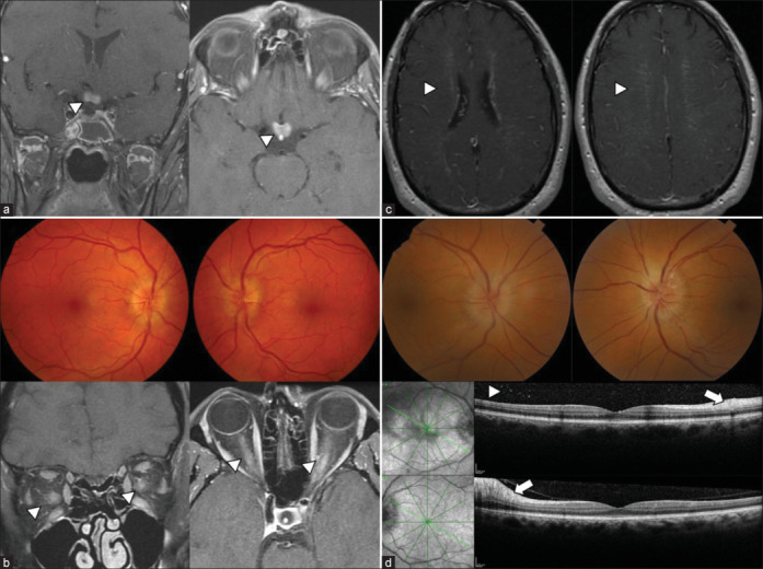
This review article represents a collaborative effort across continents, bringing together the latest developments in neuro-ophthalmology with a focus on innovative diagnostic and therapeutic modalities that are shaping the future of the field. Among the most significant advancements is the rise of optical coherence tomography (OCT), now recognized as an indispensable tool in neuro-ophthalmological research, providing unparalleled insights into optic nerve and central nervous system pathologies. Gene therapy, particularly for conditions such as Leber's hereditary optic neuropathy, marks a new frontier in personalized medicine, offering hope for previously untreatable conditions. The article also examines the transformative role of telemedicine and artificial intelligence (AI) in clinical practice, which are revolutionizing patient care and enhancing diagnostic precision. Furthermore, it highlights the impact of novel serological biomarkers on the understanding and management of immune-mediated optic neuritis, and discusses the introduction of new therapeutic agents like Tocilizumab and Teprotumumab, which are redefining treatment paradigms. Collectively, these advancements reflect the profound influence of modern medicine on neuro-ophthalmology, paving the way for improved patient outcomes and fostering new avenues for research and clinical practice.
Copyright © 2024 Copyright: © 2024 Indian Journal of Ophthalmology.
Conflict of interest statement
Nancy Newman: NJN is a consultant for GenSight Biologics, Santhera/Chiesi, Stoke, Neurophoenix, Neurophth and Avidity; received research support from GenSight Biologics and Santhera/Chiesi; and was a participant in educational webinars sponsored by WebMD Global Medscape and First Class/. John J Chen:JJC is a consultant to UCB and Amgen. Susan Mollan: SPM reports consultancy fees (Invex Therapeutics, Velux foundation), advisory board fees (Invex Therapeutics, Gensight, Ocular therapeutix), and speaker fees (Heidelberg engineering, Chugai Roche Ltd, Allergan, Santen, Teva UK, Chiesi, and Santhera). Dan Milea :DM is an advisory board member of Optomed, Finland.
Figures


References
-
- O. C. T. Sub-Study Committee for NORDIC Idiopathic Intracranial Hypertension Study Group ; Auinger P, Durbin M, Feldon S, Garvin M, Kardon R, et al. Baseline OCT measurements in the idiopathic intracranial hypertension treatment trial, part I: Quality control, comparisons, and variability. Invest Ophthalmol Vis Sci. 2014;55:8180–8. - PMC - PubMed
-
- Kaya Tutar N, Kale N. The relationship between lumbar puncture opening pressure and retinal nerve fiber layer thickness in the diagnosis of idiopathic intracranial hypertension: Is a lumbar puncture always necessary? Neurologist. 2024;29:91–5. - PubMed
Publication types
MeSH terms
Grants and funding
LinkOut - more resources
Full Text Sources
