. 2024 Dec;15(1):2421983.
doi: 10.1080/21505594.2024.2421983. Epub 2024 Nov 4.

The arginine/ornithine binding protein ArgT plays an essential role in Brucella neotomae/ Brucella melitensis to prevent intracellular killing and contribute to chronic persistence in the host

Affiliations

The arginine/ornithine binding protein ArgT plays an essential role in Brucella neotomae/ Brucella melitensis to prevent intracellular killing and contribute to chronic persistence in the host

Sushree Rekha Mallik et al. Virulence. 2024 Dec.

Abstract

Brucella species are facultative intracellular bacterial pathogens that cause the contagious zoonotic disease, brucellosis. Brucella spp. infect a wide range of animals, including livestock, wild animals, and marine mammals. Compared with other invasive bacterial pathogens, partial information is available on the virulence factors of Brucella that enable them to survive in the host. Here, we performed transposon-based random mutagenesis of B. neotomae and identified the arginine/ornithine binding protein, ArgT, as one of the crucial virulence determinants of Brucella. Deleting ArgT from B. neotomae or B. melitensis resulted in its attenuation in macrophages, which was restored upon complementation with an ArgT expression plasmid. We observed that macrophages infected with ΔArgT-B. neotomae produced elevated levels of NO due to the inability of these mutants to deplete the host intracellular arginine through their importer. Furthermore, defective survival of ΔArgT B. neotomae and B. melitensis was observed in the infected mice, which correlated with enhanced NO production in the mice. Our studies revealed that ArgT plays a vital role in preventing intracellular killing and contributes to the chronic persistence of B. neotomae/B. melitensis in the host. This study highlights the essential role of arginine in clearing intracellular infections and the subversion of this host defense mechanism by intracellular pathogens for their chronic persistence.

Keywords: ArgT; Brucella; arginine; attenuation; iNOS; nitric oxide.


Figures

Figure 1.
Figure 1.
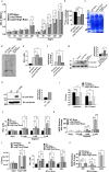
(a) ArgT-B. neotomae mutant exhibits defective intracellular survival in the macrophages. RAW264.7 cells were infected with wild-type (WT-Bneo) or Transposon-insertional mutant of B. neotomae (C13-Bneo), followed by quantification of the intracellular load of bacteria by enumeration of CFU at indicated times post-infection. (b) Fluorescence microscopy images showing the intracellular load of WT-Bneo or C13-Bneo expressing GFP. RAW264.7 cells were infected with GFP-expressing Brucella, followed by analyzing the infected cells using a fluorescent microscope at 24 hours post-infection. The cell nuclei were stained with DAPI (blue). Scale bar, 50 µm. The images are representative of three independent experiments where 12 fields were captured and analyzed for each experiment. The right panel indicates the quantification of the number of intracellular bacteria using ImageJ software. (c) Macrophage infection studies using WT (WT-Bneo) or ΔArgT-B. neotomaeArgT-Bneo) or ΔArgT-B. neotomae complemented with ArgT expression plasmid (ΔArgT+ArgT-Bneo). RAW264.7 cells were infected with indicated strains of B. neotomae, followed by the enumeration of CFUs at various times post-infection. (d) The intracellular load of WT (WT-Bmel) or ΔArgT B. melitensisArgT-Bmel) or ΔArgT+ArgT B. melitensisArgT+ArgT-Bmel) in RAW264.7 cells at indicated times post-infection. The data are presented as the mean ± SEM from at least three independent experiments (***, p < 0.001).
Figure 2.
Figure 2.
(a) ΔArgT-B. neotomae exhibits a defective arginine uptake. Growth dynamics of WT (WT-Bneo) or ΔArgT-B. neotomaeArgT-Bneo) or ΔArgT+ArgT-B. neotomaeArgT+ArgT-Bneo) in the DMEM medium with or without arginine. The indicated B. neotomae strains were inoculated into the arginine (-) DMEM or arginine (-) DMEM supplemented with L-arginine. The OD was analyzed at indicated times post-inoculation. (b) The uptake of C14-labeled L-arginine by wild-type or mutant strains of B. neotomae. The uptake assay was performed with WT-Bneo or ΔArgT-Bneo or ΔArgT+ArgT-Bneo in the presence of 14C(U) L-arginine. subsequently, the bacteria were lysed, and the imported 14C(U) L-arginine was quantified using the liquid scintillation counter .(c-d) Analysis of incorporated 14C(U) L-arginine in the Brucella proteins. After the 14C(U) L-arginine uptake assay, WT-Bneo or ΔArgT-Bneo or ΔArgT+ArgT-Bneo were cultured for 24 h, followed by lysing the cells and resolving the equal amount of total protein by SDS-PAGE (c). The gel was then dried and analyzed using a phosphor-imager (d). (e) ΔArgT-Bneo-infected macrophages produce elevated levels of NO. RAW264.7 cells were infected with WT-Bneo or ΔArgT-Bneo or ΔArgT+ArgT-Bneo, followed by quantification of NO levels at 24 h post-infection. (f-g) enhanced level of iNOS expression in the macrophages infected with ΔArgT-Bneo. RAW264.7 cells were infected with WT-Bneo or ΔArgT-Bneo or ΔArgT+ArgT-Bneo, followed by quantification of iNOS mRNA levels by qPCR (f) and iNOS protein levels by immunoblotting (g) at 24 h post-infection. The membranes were probed with anti-iNOS antibody to detect the endogenous levels of iNOS. Actin served as the loading control. The right panel indicates the densitometry of iNOS bands normalized with actin. The immunoblot is representative of three different experiments. (h) Treatment of macrophage with IFN-γ induces iNOS expression. RAW264.7 cells were treated with IFN-γ for 24 h, followed by evaluating the iNOS level by immunoblotting. The membrane was probed with the anti-iNOS antibody to detect the endogenous levels of iNOS. Actin served as the loading control. The right panel indicates the densitometry of iNOS bands normalized with actin. The immunoblot is representative of two independent experiments. (i) WT or ΔArgT-B. neotomae exhibits defective survival in the IFN-γ-treated macrophages. RAW264.7 cells were treated with IFN-γ for 24 hours, followed by infection with WT or ΔArgT or ΔArgT+ArgT-Bneo. Twenty-four hours post-infection, CFU was enumerated. (j-l) The addition of L-arginine enhanced host-induced NO and iNOS levels in macrophages. RAW264.7 cells were infected with WT-Bneo or ΔArgT-Bneo or ΔArgT+ArgT-Bneo in (-) arginine DMEM or arginine (-) DMEM supplemented with indicated concentration of L-arginine, followed by quantification of NO levels in the culture supernatants (j), iNOS expression levels in the cells (k), and determination of the intracellular load of B. neotomae strains at 24 h post-infection (l). (m-n) the levels of NO and iNOS in the macrophages infected with various MOIs of Brucella. RAW264.7 cells were infected with an increasing MOI of WT-Bneo or ΔArgT-Bneo or ΔArgT+ArgT-Bneo, followed by quantification of NO levels in the culture supernatants (m) and iNOS expression levels in the cells (n) at 24 h post-infection. The data are presented as the mean ± SEM from at least three independent experiments (*, p < 0.05; **, p < 0.01; ***, p < 0.001; ****, p < 0.0001).
Figure 3.
Figure 3.
(a-b) expression levels of ArgT and rocF in B. neotomae at various time points of infection. RAW264.7 cells were infected with WT-B. neotomae, followed by harvesting the infected cells at indicated time points and quantification of expression levels of ArgT (a) and rocF (b) of B. neotomae by qPCR. (c-d) expression levels of ArgT and rocF in B. neotomae with increasing concentrations of arginine. RAW264.7 cells were infected with WT-B. neotomae in DMEM (-) arginine or DMEM (-) arginine supplemented with indicated concentrations of L-arginine for 24 h, followed by quantification of ArgT (c) and rocF (d) expression levels by qPCR. The qPCR data were normalized with 16s rRNA, and relative mRNA expression was quantified with respect to uninfected macrophages for the corresponding time point of infection. (e) Upregulation of cationic amino acid transporter in the Brucella-infected macrophages. RAW264.7 macrophages were infected with WT-B. neotomae (WT-Bneo), followed by quantification of the expression levels of CAT2b at indicated time points by qPCR. The data were normalized with GAPDH, and relative mRNA expression was determined with respect to the uninfected macrophages for each time point of infection. The data are presented as the mean ± SEM from at least three independent experiments (*, p < 0.05; **, p < 0.01; ***, p < 0.001; ****, p < 0.0001).
Figure 4.
Figure 4.
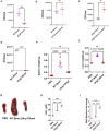
(a) ΔArgT-B. neotomae exhibits an attenuated phenotype in mice. Six to eight weeks old BALB/c mice were infected with WT (WT-Bneo) or ΔArgT-B. neotomaeArgT-Bneo). Fourteen days post-infection, the mice were sacrificed, followed by the collection of spleens. The spleens were homogenized, followed by an enumeration of CFU to determine the splenic load of WT or ΔArgT-Bneo. (b-c) ΔArgT-B. neotomae shows enhanced survival in the mice treated with iNOS inhibitor. BALB/c mice infected with WT-Bneo or ΔArgT-Bneo for 10 days followed by treatment with the iNOS inhibitor for 3 days. Mice were sacrificed on the 14th day, followed by the enumeration of the splenic load of Brucella. (d)ΔArgT-B. melitensis presents an attenuated phenotype in mice. Six to eight weeks old BALB/c mice were infected with WT (WT-Bmel) or ΔArgT-B. melitensisArgT-Bmel). fourteen days post-infection, the mice were sacrificed, followed by the collection of blood, spleens, and liver. The spleens were homogenized, followed by an enumeration of CFU to determine the splenic load of WT or ΔArgT-Bmel. (e-g) the weights of the spleen (e) and liver (f) of mice infected with WT or ΔArgT-Bmel. (g) The photograph showing the spleen of mice infected with WT or ΔArgT-Bmel. PBS was used as the negative control. (h) Levels of NO and iNOS (I) in the mice infected with WT or ΔArgT-Bmel. The NO levels were determined in the serum of infected mice using Griess reagent, and iNOS expression levels were examined in the spleen by qPCR analysis. The data were normalized with GAPDH, and relative mRNA expression was determined with respect to the group treated with PBS. The data are presented as the mean ± SEM from at least two independent experiments (p < 0.01; ***, p < 0.001; ****, p < 0.0001).

References

    1. Gwida M, Al Dahouk S, Melzer F, et al. Brucellosis – regionally emerging zoonotic disease? Croat Med J. 2010;51(4):289–16. doi: 10.3325/cmj.2010.51.289 - DOI - PMC - PubMed
    1. Franc KA, Krecek RC, Hasler BN, et al. Brucellosis remains a neglected disease in the developing world: a call for interdisciplinary action. BMC Public Health. 2018;18(1):125. doi: 10.1186/s12889-017-5016-y - DOI - PMC - PubMed
    1. Nicoletti P. Brucellosis: past, present and future. Prilozi. 2010;31(1):21–32. - PubMed
    1. Khan MZ, Zahoor M. An overview of brucellosis in cattle and humans, and its serological and molecular diagnosis in control strategies. Trop Med Infect Dis. 2018;3(2):65. doi: 10.3390/tropicalmed3020065 - DOI - PMC - PubMed
    1. Corbel MJ. Brucellosis: an overview. Emerg Infect Dis. 1997;3(2):213–221. doi: 10.3201/eid0302.970219 - DOI - PMC - PubMed

Publication types

MeSH terms

LinkOut - more resources