. 2024 Oct 23;57(2):223-236.
doi: 10.3724/abbs.2024185.

CircMALAT1 promotes the proliferation and metastasis of intrahepatic cholangiocarcinoma via the miR-512-5p/VCAM1 axis

Affiliations

CircMALAT1 promotes the proliferation and metastasis of intrahepatic cholangiocarcinoma via the miR-512-5p/VCAM1 axis

Meixia Zhang et al. Acta Biochim Biophys Sin (Shanghai). .

Abstract

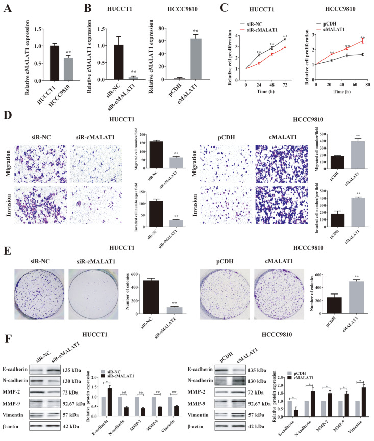
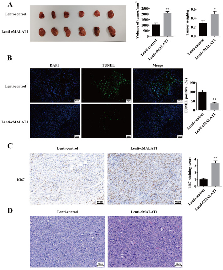
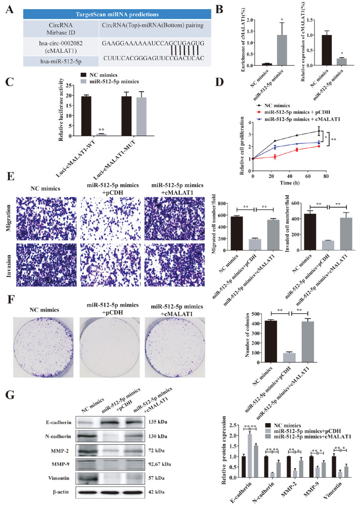
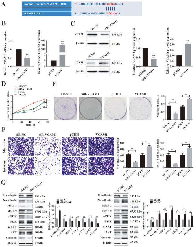
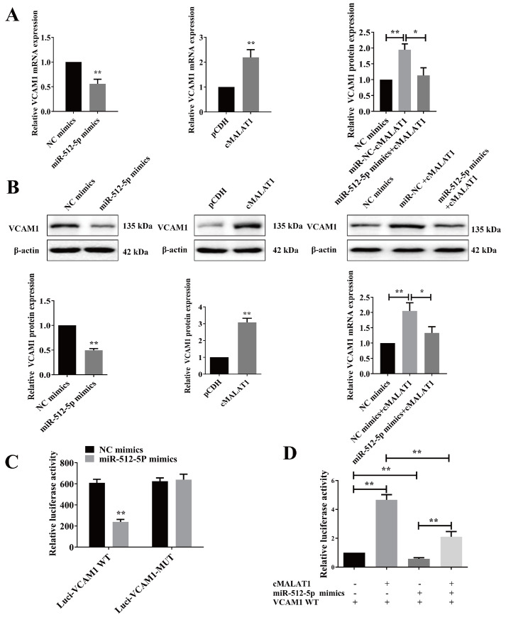
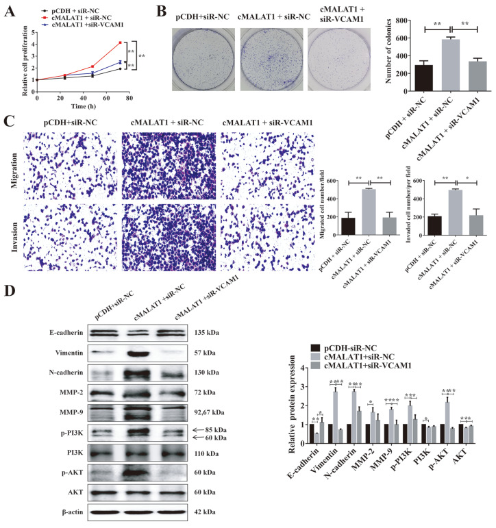
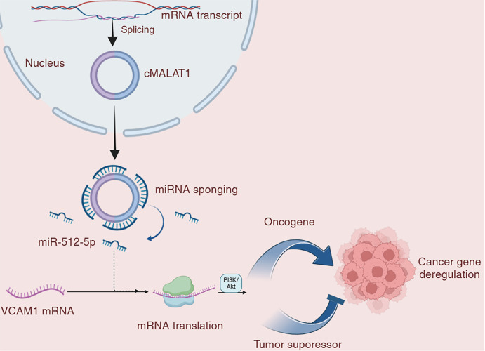
Circular RNAs play a pivotal role in the progression of various cancers. In our previous study, we observed high expression of the circRNA MALAT1 (cMALAT1) in intrahepatic cholangiocarcinoma (ICC) cells co-incubated with activated hepatic stellate cells. This study is designed to explore the roles of cMALAT1 and the underlying mechanisms in ICC. We find that cMALAT1 significantly facilitates the progression of ICC both in vitro and in vivo. The binding between cMALAT1 and miR-512-5p is subsequently confirmed through RNA pull-down experiments. As anticipated, the application of miR-512-5p mimics noticeably reverses the cMALAT1 overexpression-induced malignant phenotypes of ICC cells. Furthermore, VCAM1 is identified as a downstream gene of the cMALAT1/miR-512-5p axis. Importantly, silencing of VCAM1 not only effectively suppresses the malignant phenotypes of ICC cells but also significantly impairs the functions of cMALAT1. Our study reveals that cMALAT1 promotes the progression of ICC by competitively binding to VCAM1 mRNA with miR-512-5p, leading to the upregulation of VCAM1 expression and the activation of the PI3K/AKT signaling pathway.

Keywords: EMT; ICC; VCAM1; circular MALAT1; miR-512-5p.


Conflict of interest statement

The authors declare that they have no conflict of interest.

Figures

None
Figure 1
cMALAT1 regulates the malignant phenotype of ICC in HCCC9810 and HUCCT1 cells (A,B) The relative cMALAT1 expression in cells (A) and the knockdown or overexpression efficiency of cMALAT1 in cells (B) were tested via qRT-PCR. (C–E) Proliferation (C), migration, invasion (D), and colony formation (E) abilities were examined via MTT, transwell, and colony formation assays, respectively. (F) The expressions of MMP2, MMP9, and EMT-related proteins were also examined via western blot analysis. All the data were obtained from at least three independent experiments, and shown as the mean ± SD. *P < 0.05, **P < 0.01.
None
Figure 2
cMALAT1 promotes the malignant phenotype of ICC in xenograft tumors Xenografts were established by subcutaneously injecting 5×10⁶ HCCC9810 cells stably expressing cMALAT1 (Lenti-cMALAT1-OE, n = 6) or control cells (Lenti-control, n = 6) into the right flank of nude mice. (A) The volume and weight of the tumor tissues were measured. (B,C) Apoptosis (B) and the expression of Ki67 (C) in tumor tissues were examined by TUNEL and IHC assays, respectively. (D) HE staining of tumor tissue. All the data were obtained from at least three independent experiments, and shown as the mean ± SD. *P < 0.05, **P < 0.01.
None
Figure 3
The binding of miR-512-5p to cMALAT1 reverses its pro-malignant effect on ICC cells (A) The miRNAs that bind to cMALAT1 were predicted via the circular RNA interactome. (B) The enrichment of cMALAT1 was examined through a biotin-coupled miRNA pull-down assay (left panel), and the expression of cMALAT1 in the supernatant samples after the biotin-coupled miRNA pull-down assay was detected via qPCR (right panel) to verify the binding of miR-512-5p to cMALAT1. (C) The binding site identified in cMALAT1 was mutated to perform a dual-luciferase reporter assay. (D–F) Cell proliferation (D), migration, invasion (E), and colony formation (F) abilities of HCCC9810 cells were examined via MTT, transwell, and colony formation assays, respectively. (G) The expressions of EMT-related proteins was examined via western blot analysis. All the data were obtained from at least three independent experiments, and shown as the mean ± SD. *P < 0.05, **P < 0.01.
None
Figure 4
VCAM1 plays a pro-cancer role in HCCC9810 cells (A) The TargetScan Human database (https://www.targetscan.org/vert_80/) was used to predict the binding sites between miR-512-5p and VCAM1. (B,C) The effects of VCAM1 overexpression and siR-VCAM1 in HCCC9810 cells were verified by PCR (B) and WB (C) experiments. (D–F) The proliferation (D), colony formation (E), invasion, and migration (F) abilities of HCCC9810 cells were examined via MTT, colony formation, and transwell assays, respectively. (G) WB was used to verify the expression of EMT and key PI3K/AKT molecules in the siR-VCAM1- and VCAM1-overexpressing groups. All the data were obtained from at least three independent experiments, and shown as the mean ± SD. *P < 0.05, **P < 0.01.
None
Figure 5
VCAM1 is regulated by cMALAT1 and miR-512-5p mimics (A,B) The expression of VCAM1 was examined by PCR (A) and western blot analysis (B). (C) A dual-luciferase reporter assay was performed to examine the impact of miR-512-5p on VCAM1-Wt and VCAM1-Mt. (D) Luciferase activity was detected in miR-512-5p mimic HCCC9810 cells co-transfected with cMALAT1-overexpressing and VCAM1-Wt-overexpressing plasmids. All the data were obtained from at least three independent experiments, and shown as the mean ± SD. *P < 0.05, **P < 0.01.
None
Figure 6
VCAM1 promotes the malignant phenotype of ICC in HCCC9810 cells (A–C) Proliferation (A), colony formation, (B) migration, and invasion (C) abilities were examined via MTT, colony formation, and transwell assays, respectively. (D) The expressions of MMP2, MMP9, p-P13K, p-AKT, and EMT-related proteins were also examined via western blot analysis. All the data were obtained from at least three independent experiments, and shown as the mean ± SD. *P < 0.05, **P < 0.01.
None
Figure 7
A novel mechanism by which the interaction of the cMALAT1/miR-512-5p/VCAM1 network regulates the cancer progression of ICC

Similar articles

Cited by

References

    1. Siegel RL, Miller KD, Fuchs HE, Jemal A. Cancer statistics, 2022. CA Cancer J Clin. . 2022;72:7–33. doi: 10.3322/caac.21708. - DOI - PubMed
    1. Beal EW, Tumin D, Moris D, Zhang XF, Chakedis J, Dilhoff M, Schmidt CM, et al. Cohort contributions to trends in the incidence and mortality of intrahepatic cholangiocarcinoma. Hepatobiliary Surg Nutr. . 2018;7:270–276. doi: 10.21037/hbsn.2018.03.16. - DOI - PMC - PubMed
    1. Sabbatino F, Villani V, Yearley JH, Deshpande V, Cai L, Konstantinidis IT, Moon C, et al. PD-L1 and HLA class I antigen expression and clinical course of the disease in intrahepatic cholangiocarcinoma. Clin Cancer Res. . 2016;22:470–478. doi: 10.1158/1078-0432.CCR-15-0715. - DOI - PMC - PubMed
    1. Xia X, Li X, Li F, Wu X, Zhang M, Zhou H, Huang N, et al. A novel tumor suppressor protein encoded by circular AKT3 RNA inhibits glioblastoma tumorigenicity by competing with active phosphoinositide-dependent Kinase-1. Mol Cancer. . 2019;18:131. doi: 10.1186/s12943-019-1056-5. - DOI - PMC - PubMed
    1. Wang L, Long H, Zheng Q, Bo X, Xiao X, Li B. Circular RNA circRHOT1 promotes hepatocellular carcinoma progression by initiation of NR2F6 expression. Mol Cancer. . 2019;18:119. doi: 10.1186/s12943-019-1046-7. - DOI - PMC - PubMed

MeSH terms