Case Reports
. 2024 Sep 25;16(9):e70193.
doi: 10.7759/cureus.70193. eCollection 2024 Sep.

Cortical Venting: A Simple Surgical Adjunct for the Treatment of Long Bone Nonunion

Affiliations
Case Reports

Cortical Venting: A Simple Surgical Adjunct for the Treatment of Long Bone Nonunion

Sean M Wade et al. Cureus. .

Abstract

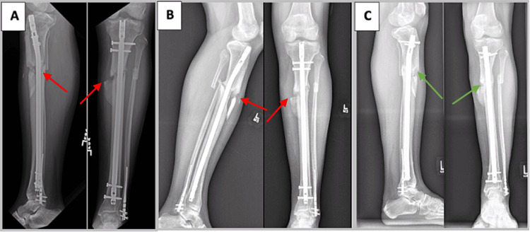
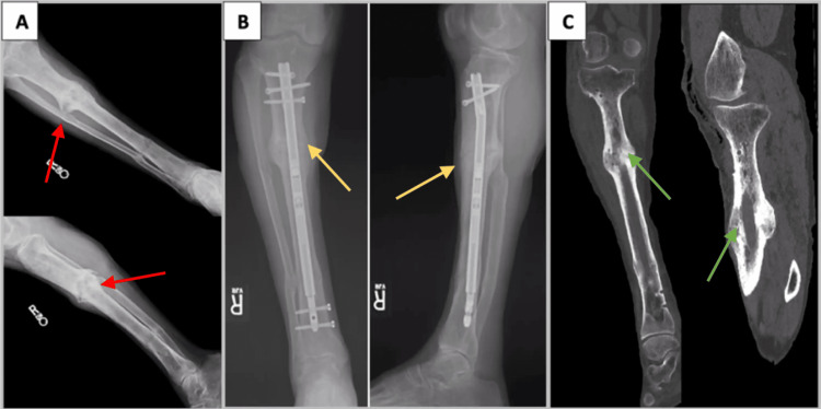
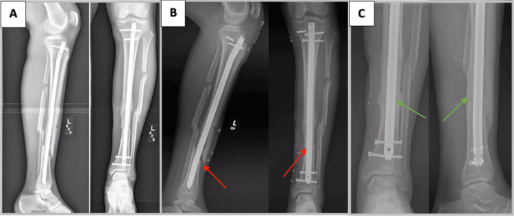
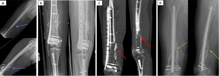
Nonunions are a vexing problem for the orthopedic surgeon. Herein, we describe an adjunct to the standard exchange nailing procedure adapted from an established limb lengthening technique in which cortical vents are drilled adjacent to the nonunion site. These transcortical drill tunnels facilitate local dispersion of the osteogenic intramedullary reamings around the nonunion site during the exchange nailing, whereby the extruded reamings serve as autograft for the nonunion. This simple adjunctive technique can increase the likelihood of achieving union when performed with an exchange nailing procedure as demonstrated by our case series of recalcitrant tibia and femoral nonunions successfully treated when this surgical adjunct was implemented.

Keywords: cortical venting; exchange nailing; long bone fractures; nonunion; orthopaedic trauma.


Conflict of interest statement

Human subjects: Consent was obtained or waived by all participants in this study. Conflicts of interest: In compliance with the ICMJE uniform disclosure form, all authors declare the following: Payment/services info: All authors have declared that no financial support was received from any organization for the submitted work. Financial relationships: All authors have declared that they have no financial relationships at present or within the previous three years with any organizations that might have an interest in the submitted work. Other relationships: All authors have declared that there are no other relationships or activities that could appear to have influenced the submitted work.

Figures

Figure 1
Figure 1. A 30-year-old man with oligotrophic tibial nonunion (case 1)
A) Radiographs of the patient’s left leg taken eight months after injury demonstrating a nonunited (red arrows) and deformed tibia. B) Persistent nonunion (red arrows) 12 months following the patient’s second exchange nailing and allogeneic bone grafting. At this time, priority was placed on achieving union through a third exchange nailing rather than concomitantly addressing the tibia’s residual deformity. C) Tibial union (green arrows) is evident on radiographs taken one year after performing our modified exchange nailing operation utilizing cortical vent tunnels. The patient has since declined to proceed with tibial malunion correction surgery.
Figure 2
Figure 2. A 69-year-old male smoker with a hypertrophic tibial nonunion (case 2)
A) Radiographs after failed Ilizarov external fixation treatment complicated by osteomyelitis demonstrating a persistent hypertrophic nonunion (red arrows). B) Approximately one year after exchange nailing with cortical venting and insertion of a telescoping motorized intramedullary nail in which bridging callous formation (yellow arrows) is evident. C) CT of the tibia at 18 months after modified exchanged nailing procedure and subsequent intramedullary nail removal demonstrating union (green arrows).
Figure 3
Figure 3. A 56-year-old male smoker with oligotrophic nonunion of a segmental tibial fracture (case 3)
A) Radiographs of the patient’s left tibia four months after injury, status post tibia and fibula intramedullary nailing at an outside hospital with soleus flap coverage and skin grafting. B) Radiographs of the left tibia demonstrating persistent nonunion (red arrows) with no callous formation at fracture site six months after index surgery. C) Radiographs demonstrating union (green arrows) approximately one year after exchange nailing performed with adjunctive cortical vent drill tunnels. The patient was pain-free at the former tibial nonunion site.
Figure 4
Figure 4. A 32-year-old man with an open, comminuted distal femur fracture that developed into an oligotrophic nonunion (case 4)
A) Radiographs four weeks status post retrograde nailing for the comminuted distal third femur fracture (blue arrows) at an outside institution, with approximately two centimeters of shortening and no evidence of callus formation at the fracture site. Ipsilateral tibial nailing was performed at the time of initial retrograde femoral nailing for an open tibia fracture. B) Immediate postoperative imaging status post femoral nail removal with revision to lateral distal femur locking plate and bone grafting. C) CT scan eight months after femoral plating and bone grafting operation demonstrating a persistent nonunion (red arrows). D) Radiographs nine months after exchange nailing with pre-drilled cortical vent tunnels demonstrating robust bridging bone formation (green arrows) at the former nonunion site.

References

    1. Tibia shaft fractures: costly burden of nonunions. Antonova E, Le TK, Burge R, Mershon J. BMC Musculoskelet Disord. 2013;14:42. - PMC - PubMed
    1. Treatment of severely open tibial fractures, non-unions, and fracture-related infections with a gentamicin-coated tibial nail-clinical outcomes including quality of life analysis and psychological ICD-10-based symptom rating. Walter N, Popp D, Freigang V, Nerlich M, Alt V, Rupp M. J Orthop Surg Res. 2021;16:270. - PMC - PubMed
    1. Outcomes of patients with nonunion after open tibial shaft fractures with or without soft tissue coverage procedures. Egol KA, Littlefield CP, Walden T, Leucht P, Levine J, Konda S. J Orthop Trauma. 2021;35:0–6. - PubMed
    1. Path analysis of factors for delayed healing and nonunion in 416 operatively treated tibial shaft fractures. Audigé L, Griffin D, Bhandari M, Kellam J, Rüedi TP. Clin Orthop Relat Res. 2005;438:221–232. - PubMed
    1. Fractures of the tibia: can their outcome be predicted? Gaston P, Will E, Elton RA, McQueen MM, Court-Brown CM. J Bone Joint Surg Br. 1999;81:71–76. - PubMed

Publication types

LinkOut - more resources