

Case Report: Esophageal squamous cell carcinoma in a 13-year-old boy with a history of esophageal atresia with tracheoesophageal fistula
- PMID: 39463732
- PMCID: PMC11502401
- DOI: 10.3389/fped.2024.1438242
Case Report: Esophageal squamous cell carcinoma in a 13-year-old boy with a history of esophageal atresia with tracheoesophageal fistula
Abstract
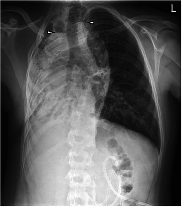
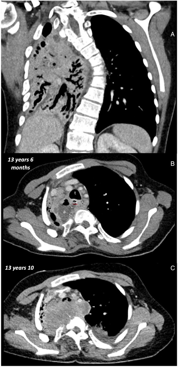
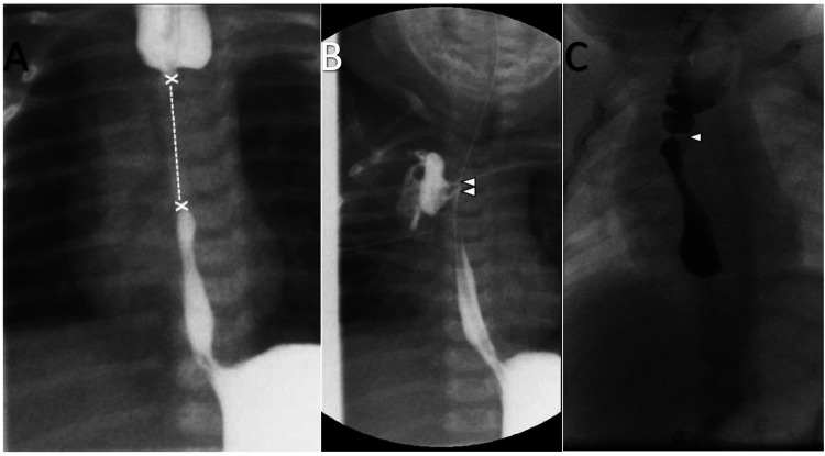
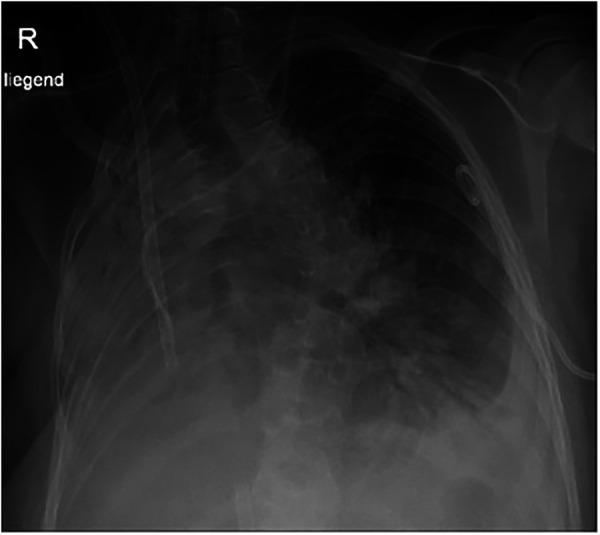
In adults, esophageal cancers are a global health concern. Esophageal squamous cell carcinoma (ESCC) accounts for approximately 90% of esophageal carcinomas. The prognosis of esophageal cancers remains dismal, with a five-year survival rate below 20%. It typically affects older patients, and for now, ESCC after esophageal atresia has not been reported in patients younger than 18 years. We present an exceptional case of an ESCC in a 13-year-old boy with a history of esophageal atresia and corrective surgery in infancy. After the surgery the patient was lost to surgical follow up for over ten years and then presented to our emergency department with respiratory distress requiring antibiotic therapy and supplemental oxygen. Radiologic imaging revealed a volume reduction of the right lung with bronchiectasis, as well as esophageal stenosis at the level of the previous anastomosis, with an adjacent abscess in the right lung. These changes may have arisen due to a chronic fistula from the esophagus to the right lung. Initial interventional therapy with a stent implantation had no lasting success and, in an effort to prevent further aspiration into the right lung, a cervical esophagus stoma was established, and the patient received prolonged antibiotic treatment. However, a thoracic CT scan performed 4 months later revealed a large, retrospectively progressive prevertebral mass originating from the distal portion of the esophagus below the stenosis, compressing the trachea and the right main bronchus. The patient's condition rapidly worsened and he developed respiratory failure, requiring veno-venous extracorporeal membrane oxygenation. Unfortunately, an endoscopic biopsy revealed an advanced ESCC. With no rational treatment options available, we changed the goals of care to a palliative setting. The key message of this case is that in adolescents with chronic infections, an abscess can potentially mask a malignant transformation. Therefore, in adolescents, with an history of corrective surgery for esophageal atresia and chronic complications, consideration should also be given to the possibility of squamous cell carcinoma of the esophagus.
Keywords: ESCC (esophageal squamous cell carcinoma); cancer; child—age; esophageal atresia; esophageal repair.
© 2024 Bernar, Mayerhofer, Fuchs, Schweigmann, Gassner, Crazzolara, Hetzer, Klingkowski, Zschocke and Cortina.
Conflict of interest statement
The authors declare that the research was conducted in the absence of any commercial or financial relationships that could be construed as a potential conflict of interest.
Figures
References
Publication types
LinkOut - more resources
Full Text Sources