Case Reports
. 2024 Oct 19:2024:2594659.
doi: 10.1155/2024/2594659. eCollection 2024.

Surgical Management of Complex Multiligament Knee Injury: Case Report

Affiliations
Case Reports

Surgical Management of Complex Multiligament Knee Injury: Case Report

Ramon Alonso Prieto Baeza et al. Case Rep Orthop. .

Abstract

Multiligament knee injuries (MLKIs) frequently require immediate intervention to prevent severe complications, including vascular injury. We present the case of a 51-year-old male who sustained a traumatic right knee dislocation following a motor vehicle accident. The patient exhibited significant tibiofemoral dissociation with Grade 3 instability, classified as Schenck KD IV. Immediate reduction and external fixation were performed, followed by definitive surgical management, which included fibular sling, MPFL and MCL repair, and double-bundle and double-tunnel ACL and PCL reconstruction with looped proximal tibial fixation. The patient showed an excellent early postoperative outcome, with minimal edema, manageable moderate pain, and a full range of motion by the 30-day follow-up. This case underscores the effectiveness of combining fibular sling, MPFL, and MCL, with anatomical double-bundle ACL and PCL reconstruction in the treatment of complex MLKIs. The level of evidence is IV.

Keywords: anatomical reconstruction; double-bundle technique; knee stability; multiligament knee injury.


Conflict of interest statement

The authors declare no conflicts of interest.

Figures

Figure 1
Figure 1
(a, b) Axial, (c, d) AP, and (e) lateral CT-scan: dislocation of the femorotibial joint is observed, with medial displacement of the femoral portion. Additionally, an avulsion fracture is identified on the lateral tibial surface, specifically in its medial portion. Furthermore, lateral displacement of the patella is noted. The quadriceps and patellar tendons exhibit normal density.
Figure 2
Figure 2
Angiotomography results. (a, b) Axial and (c, d) lateral view: the common femoral vein, superficial femoral vein, and deep femoral vein are intact.
Figure 3
Figure 3
Physical examination performed on anesthetized patient prior to external fixation involving a combination of rotational and valgus forces, making it sensitive to both rotational and anterior-posterior stability, with a primary plane of motion in the transverse plane. Pivot shift maneuver observed from left to right is presented, with clear tibiofemoral dissociation. The PSM app was utilized to calculate the degree of instability using quantitative data, yielding a result of Grade 3 instability.
Figure 4
Figure 4
X-ray: (a) AP and (b) lateral of the right knee post external fixation.
Figure 5
Figure 5
CT scan following multiligamentous reconstruction demonstrates adequate reduction of dislocation. (a–c) tunnel trajectory is identified as coursing caudocephalically toward the lateral femoral condyle, with an oblique disposition. Additionally, there is discreetly oblique cephalocaudal tunneling of the medial femoral condyle, reaching the inner portion of the said condyle. (d, e) Oblique tunneling of the internal tibial plateau is noted, with bone fragments within; slightly medial to this trajectory, a second tunnel is partially occupied by a screw. (f) At the level of the proximal epiphysis of the fibula, complete anteroposterior tunneling is identified, partially occupied by a fixation screw.
Figure 6
Figure 6
The proximal (a–d) tibiofibular and femorotibial and (e) patellofemoral joints show adequate congruence without alterations in the width of their joint spaces.
Figure 7
Figure 7
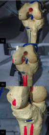
Placement of guide and brocade for the transtibial tunnel for anterior cruciate with double bundle. Bundles: (a, b) AM and (c, d) PL. The tibial tunnels are directed from the anteromedial side of the proximal metaphysis. Both lay at the same level, located 1 cm behind the anterior tibial tuberosity and 1 cm from each other. The intra-articular exit of the tunnels is arthroscopically monitored and located 8 mm anterior to the PCL, at the original ACL footprint.
Figure 8
Figure 8
Drilling of the femoral tunnels. The resident's ridge is identified as (a) the anterior boundary, with the over-the-top edge serving as (b) the posterior boundary. The femoral tunnels are then strategically positioned within the upper and lower spaces defined by these anatomical landmarks.
Figure 9
Figure 9
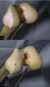
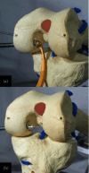
Representation in an anatomical model of (a, b) graft passage and (c, d) intraoperative image. The graft is pulled in from distal to proximal, and looped on the tibial medial proximal cortex. No fixation is performed on the tibia, and the graft should pass easily for similar lengths on both bundles. The posterolateral bundle is first fixed, and the graft is taken into maximum tension for the AM bundle fixation.
Figure 10
Figure 10
Schematic view of the double-bundle and double-tunnel ACL reconstruction with looped proximal tibial fixation procedure on a model. The U-shaped anchorage is on the tibia and the dual fixation is on the femur. This is an anatomical double-bundle double-tunnel reconstruction with no tibial hardware.
Figure 11
Figure 11
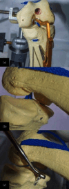
Passage of guides in the femoral condyle with a Clancy system in an (a) out–in manner, with location and exit in the footprint of the PCL at its (b) femoral insertion.
Figure 12
Figure 12
(a, b) Broaching and passage of guides through tunnels in the posterior area of the tibia. (c) 0.5 inches inferior to the articular surface with the use of a posterior guide.
Figure 13
Figure 13
(a–c) Wires are retrieved through the anterior portals for the passage of graft sutures and traction through tibial tunnels.
Figure 14
Figure 14
Graft with 25–30 cm length and 8–10 mm width per bundle. (a, b) The graft is introduced in one of the tibial tunnels and is passed subcutaneously through the tunnels in the U-shaped perforation, leaving the union in the femoral area. (c) Screw placement in tibial tunnels with knee fixation in extension.
Figure 15
Figure 15
(a, b) Tibial tunnel drilling, (c) graft passage, and (d) fixation using bioabsorbable screws.
Figure 16
Figure 16
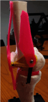
3D–printed anatomical model representation of surgical repair of MCL and MPFL.
Figure 17
Figure 17
(a) Lateral and (b) posteroanterior images of PLC repair in the anatomical model.
Figure 18
Figure 18
(a) Two months postoperative progress: The patient is walking independently without the need for support and is free of pain. (b) Physical examination demonstrates a negative anterior drawer test. However, (c) possible posterior sag may indicate some degree of residual posterior instability.

References

    1. Patel N. K., Lian J., Nickoli M., et al. Risk factors associated with complications after operative treatment of multiligament knee injury. Orthopedic Journal of Sports Medicine . 2021;9(3, article 232596712199420) doi: 10.1177/2325967121994203. - DOI - PMC - PubMed
    1. Ng J. W. G., Myint Y., Ali F. M. Management of multiligament knee injuries. EFORT Open Reviews . 2020;5(3):145–155. doi: 10.1302/2058-5241.5.190012. - DOI - PMC - PubMed
    1. Kim S. H., Park Y.-B., Kim B.-S., Lee D.-H., Pujol N. Incidence of associated lesions of multiligament knee injuries: a systematic review and meta-analysis. Orthopedic Journal of Sports Medicine . 2021;9(6, article 232596712110104) doi: 10.1177/23259671211010409. - DOI - PMC - PubMed
    1. Yasuma S., Nozaki M., Murase A., et al. Anterolateral ligament reconstruction as an augmented procedure for double-bundle anterior cruciate ligament reconstruction restores rotational stability: quantitative evaluation of the pivot shift test using an inertial sensor. The Knee . 2020;27(2):397–405. doi: 10.1016/j.knee.2020.02.015. - DOI - PubMed
    1. Berumen-Nafarrate E., Tonche-Ramos J., Carmona-González J., et al. Interpretation of the pivot test using accelerometers in the orthopedic practice. Acta Ortopédica Mexicana . 2015;29(3):176–181. - PubMed

Publication types

LinkOut - more resources