[Preprint]. 2024 Oct 17:2024.10.14.618260.
doi: 10.1101/2024.10.14.618260.

Sequential structure probing of cotranscriptional RNA folding intermediates

Affiliations

Sequential structure probing of cotranscriptional RNA folding intermediates

Courtney E Szyjka et al. bioRxiv. .

Update in

Abstract

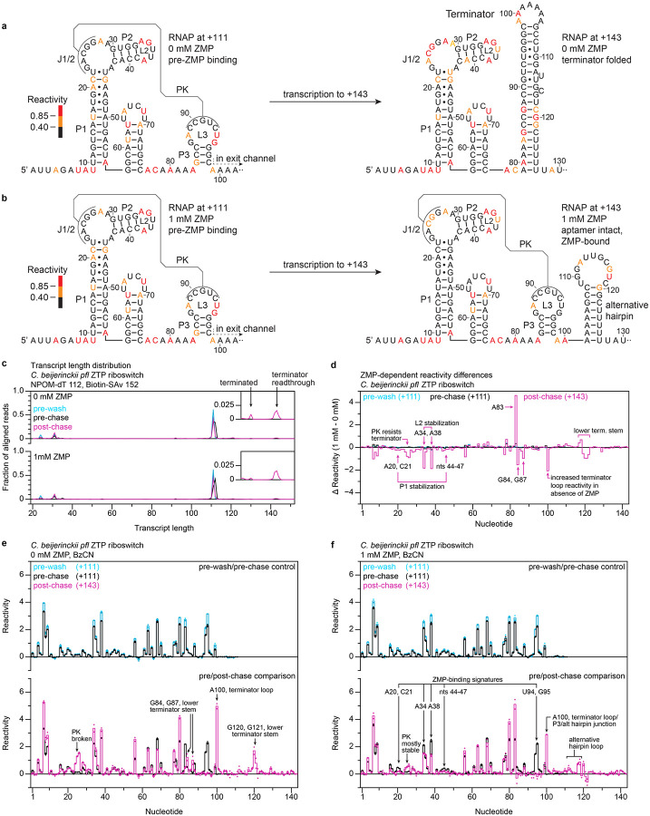
Cotranscriptional RNA folding pathways typically involve the sequential formation of folding intermediates. Existing methods for cotranscriptional RNA structure probing map the structure of nascent RNA in the context of a terminally arrested transcription elongation complex. Consequently, the rearrangement of RNA structures as nucleotides are added to the transcript can be inferred but is not assessed directly. To address this limitation, we have developed linked-multipoint Transcription Elongation Complex RNA structure probing (TECprobe-LM), which assesses the cotranscriptional rearrangement of RNA structures by sequentially positioning E. coli RNAP at two or more points within a DNA template so that nascent RNA can be chemically probed. We validated TECprobe-LM by measuring known folding events that occur within the E. coli signal recognition particle RNA, Clostridium beijerinckii pfl ZTP riboswitch, and Bacillus cereus crcB fluoride riboswitch folding pathways. Our findings establish TECprobe-LM as a strategy for detecting cotranscriptional RNA folding events directly using chemical probing.


Conflict of interest statement

Competing Interest The authors have no conflicts of interest with the contents of this article.

Figures

Figure 1.
Figure 1.. Overview of TECprobe-LM.
Single-round transcription is initiated on template DNA that contains NPOM-caged-dT and terminal biotin-streptavidin roadblocks. In the initial phase of transcription, RNAP arrests at the NPOM-caged-dT roadblock. The pre-wash sample is removed from the reaction and chemically probed, and the remaining transcription reaction is washed to remove NTPs. The NPOM cage is removed by irradiation with 365 nm UV light and the pre-chase sample is removed from the reaction and chemically probed. Upon addition of NTPs, RNAP transcribes to the terminal biotin-streptavidin roadblock and the post-chase sample is removed and chemically probed. RNAP, RNA polymerase; SRP, signal recognition particle; SAv, streptavidin; BzCN, benzoyl cyanide.
Figure 2.
Figure 2.. Cotranscriptional folding of the E. coli SRP RNA.
(a) Secondary structures of the E. coli SRP RNA folding intermediates that were assessed by TECprobe-LM colored by reactivity. Sequence within the RNAP footprint is not shown. (b) Transcript length distribution for the pre-wash, pre-chase, and post-chase samples. Traces are the average of n=2 replicates. (c) Comparison of reactivity profiles for pre-wash and pre-chase samples (upper plot) and for pre-chase and post-chase samples (lower plot). Solid lines are the average of n=2 replicates and reactivity values for individual replicates are shown as points. RNAP, RNA polymerase; PK, pseudoknot; SAv, streptavidin; BzCN, benzoyl cyanide.
Figure 3.
Figure 3.. Cotranscriptional folding of the C. beijerinckii pfl ZTP riboswitch aptamer.
(a) Secondary structures of the C. beijerinckii pfl ZTP riboswitch aptamer folding intermediates that were assessed by TECprobe-LM colored by reactivity. (b) Transcript length distribution for the pre-UV, pre-chase, and post-chase samples. Traces are the average of n=2 replicates. (c) Difference in reactivity (1 mM ZMP - 0 mM ZMP) observed for the pre-UV, pre-chase, and post-chase samples. Differences were calculated from the average reactivity values shown in (d) and (e). (d, e) Comparison of reactivity profiles for pre-UV and pre-chase samples (upper plot) and for pre-chase and post-chase samples (lower plot) in the absence (d) and presence (e) of 1 mM ZMP. Solid lines are the average of n=2 replicates and reactivity values for individual replicates are shown as points. RNAP, RNA polymerase; PK, pseudoknot; SAv, streptavidin; BzCN, benzoyl cyanide.
Figure 4.
Figure 4.. Cotranscriptional folding of the C. beijerinckii pfl ZTP riboswitch expression platform.
(a, b) Secondary structures of the C. beijerinckii pfl riboswitch expression platform folding intermediates that were assessed by TECprobe-LM in the absence (a) and presence (b) of 1 mM ZMP colored by reactivity. (c) Transcript length distribution for the pre-wash, pre-chase, and post-chase samples. Traces are the average of n=2 replicates. (d) Difference in reactivity (1 mM ZMP - 0 mM ZMP) observed for the pre-wash, pre-chase, and post-chase samples. Differences were calculated from the average reactivity values shown in (e) and (f). (e, f) Comparison of reactivity profiles for pre-wash and pre-chase samples (upper plot) and for pre-chase and post-chase samples (lower plot) in the absence (e) and presence (f) of 1 mM ZMP. Solid lines are the average of n=2 replicates and reactivity values for individual replicates are shown as points. RNAP, RNA polymerase; PK, pseudoknot; SAv, streptavidin; BzCN, benzoyl cyanide.
Figure 5.
Figure 5.. Cotranscriptional folding of the B. cereus crcB fluoride riboswitch aptamer.
(a, b) Secondary structures of the B. cereus crcB fluoride riboswitch aptamer folding intermediates that were assessed by TECprobe-LM in the absence (a) and presence (b) of 10 mM NaF colored by reactivity. Sequence within the RNAP footprint is not shown. (c) Transcript length distribution for the pre-wash, pre-chase, and post-chase samples. Traces are the average of n=2 replicates. (d) Difference in reactivity (10 mM NaF - 0 mM NaF) observed for the pre-wash, pre-chase, and post-chase samples. Differences were calculated from the average reactivity values shown in (e) and (f). (e, f) Comparison of reactivity profiles for pre-wash and pre-chase samples (upper plot) and for pre-chase and post-chase samples (lower plot) in the absence (e) and presence (f) of 10 mM NaF. Solid lines are the average of n=2 replicates and reactivity values for individual replicates are shown as points. RNAP, RNA polymerase; PK, pseudoknot; SAv, streptavidin; BzCN, benzoyl cyanide.
Figure 6.
Figure 6.. Cotranscriptional folding of the B. cereus crcB fluoride riboswitch expression platform.
(a, b) Secondary structures of the B. cereus crcB fluoride riboswitch expression platform folding intermediates that were assessed by TECprobe-LM in the absence (a) and presence (b) of 10 mM NaF colored by reactivity. Sequence within the RNAP footprint is not shown. (c) Transcript length distribution for the pre-wash, pre-chase, and post-chase samples. Traces are the average of n=2 replicates. (d) Difference in reactivity (10 mM NaF - 0 mM NaF) observed for the pre-wash, pre-chase, and post-chase samples. Differences were calculated from the average reactivity values shown in (e) and (f). (e, f) Comparison of reactivity profiles for pre-wash and pre-chase samples (upper plot) and for pre-chase and post-chase samples (lower plot) in the absence (e) and presence (f) of 10 mM NaF. Solid lines are the average of n=2 replicates and reactivity values for individual replicates are shown as points. RNAP, RNA polymerase; PK, pseudoknot; SAv, streptavidin; BzCN, benzoyl cyanide.

References

    1. Lai D., Proctor J. R. & Meyer I. M. On the importance of cotranscriptional RNA structure formation. RNA 19, 1461–1473 (2013). 10.1261/rna.037390.112 - DOI - PMC - PubMed
    1. Scharfen L. & Neugebauer K. M. Transcription Regulation Through Nascent RNA Folding. J Mol Biol 433, 166975 (2021). 10.1016/j.jmb.2021.166975 - DOI - PMC - PubMed
    1. Shine M. et al. Co-transcriptional gene regulation in eukaryotes and prokaryotes. Nat Rev Mol Cell Biol 25, 534–554 (2024). 10.1038/s41580-024-00706-2 - DOI - PMC - PubMed
    1. Al-Hashimi H. M. & Walter N. G. RNA dynamics: it is about time. Curr Opin Struct Biol 18, 321–329 (2008). 10.1016/j.sbi.2008.04.004 - DOI - PMC - PubMed
    1. Pan T. & Sosnick T. RNA folding during transcription. Annu Rev Biophys Biomol Struct 35, 161–175 (2006). 10.1146/annurev.biophys.35.040405.102053 - DOI - PubMed

Publication types

LinkOut - more resources