[Preprint]. 2024 Oct 18:2024.10.16.618588.
doi: 10.1101/2024.10.16.618588.

Methylation Clocks Do Not Predict Age or Alzheimer's Disease Risk Across Genetically Admixed Individuals

Affiliations

Methylation Clocks Do Not Predict Age or Alzheimer's Disease Risk Across Genetically Admixed Individuals

Sebastián Cruz-González et al. bioRxiv. .

Abstract

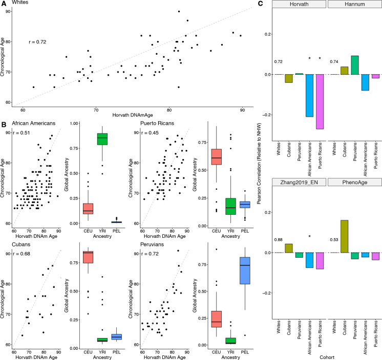
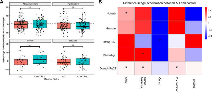
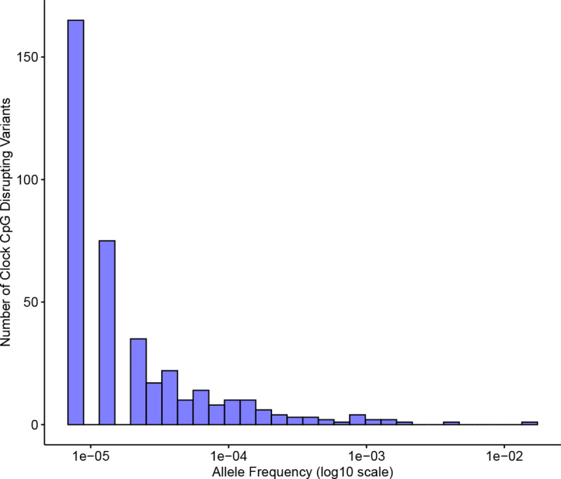
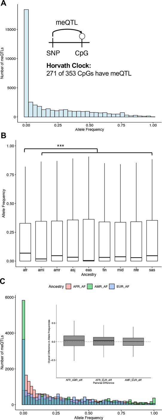
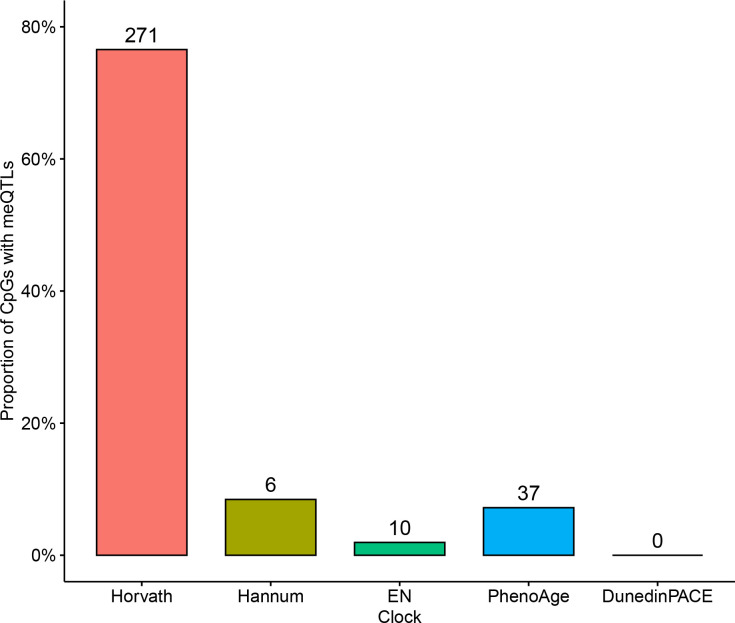
Epigenetic clocks that quantify rates of aging from DNA methylation patterns across the genome have emerged as a potential biomarker for risk of age-related diseases, like Alzheimer's disease (AD), and environmental and social stressors. However, methylation clocks have not been validated in genetically diverse cohorts. Here we evaluate a set of methylation clocks in 621 AD patients and matched controls from African American, Hispanic, and white cohorts. The clocks are less accurate at predicting age in genetically admixed individuals, especially those with substantial African ancestry, than in the white cohort. The clocks also do not consistently identify age acceleration in admixed AD cases compared to controls. Methylation QTL (meQTL) commonly influence CpGs in clocks, and these meQTL have significantly higher frequencies in African genetic ancestries. Our results demonstrate that methylation clocks often fail to predict age and AD risk beyond their training populations and suggest avenues for improving their portability.


Conflict of interest statement

4.11Competing interestsre The authors declare no competing interests.

Figures

Figure 1:
Figure 1:. Schematic of the workflow of the study.
We analyzed genome-wide methylation and genotyping data from blood samples from 621 AD and non-demented control individuals from the MAGENTA study. We applied a set of first-, second-, and third-generation methylation clocks to the individuals and estimated their genetic ancestry. This enabled us to explore the performance of methylation clocks in individuals with different genetic ancestries.
Figure 2:
Figure 2:. Methylation clock accuracy is lower in cohorts with substantial African genetic ancestry.
A: Pearson correlation between chronological age and DNAm age predicted by the Horvath clock for controls in the white MAGENTA cohort. The correlation of 0.72 is similar to previous studies of older cohorts. B: Pearson correlation between chronological age and DNAm age predicted by the Horvath clock for the genetically admixed cohorts in MAGENTA. The right plot in each pair shows the proportion of European (CEU), African (YRI), and American (PEL) global ancestry for each individual in each cohort. The two cohorts with substantial African ancestry—African Americans and Puerto Ricans—have significantly lower correlations than the other cohorts. C: Difference in correlation between chronological and predicted DNAm age for controls in each cohort compared to the white cohort controls for four methylation clocks. The baseline correlation for the white cohort controls is given in each panel; asterisks indicate a statistically significant difference from the baseline. * p < 0.05.
Figure 3:
Figure 3:. Methylation clocks rarely identify accelerated aging in admixed Alzheimer’s cohorts.
A: Comparison of the distributions of Horvath intrinsic age acceleration for AD patients and non-demented controls for each of the admixed cohorts in MAGENTA. AD patients do not show significantly higher age acceleration in any of the admixed cohorts. In contrast, the AD cases had significantly greater acceleration than controls in the white cohort (Supplementary Figure 1). NS = Not significant. B: Median differences in intrinsic age acceleration between AD patients and non-demented controls for five methylation clocks for each cohort in MAGENTA. The clocks do not consistently identfy accelerated aging in AD across cohorts, and the results also vary within cohorts. * p < 0.05.
Figure 4:
Figure 4:. Genetic variants that disrupt methylation clock CpG sites appear at extremely low frequencies.
The allele frequency distribution for single nucletide variants that disrupt one of the 353 CpG sites considered by the Horvath clock. Allele frequencies were computed across 76,156 individuals from large-scale sequencing studies harmonized in gnomAD (version 3.0).
Figure 5:
Figure 5:. Common meQTLs affect most Horvath clock CpGs and vary in frequency across ancestries.
A: The allele frequency distribution of the 29,033 unique variants associated with methylation levels at Horvath clock CpGs. Allele frequencies were computed over the 76,215 genomes in gnomAD version 4.1. Inset: Out of the 353 CpGs in the Horvath clock, 271 (77%) have at least one meQTL, i.e., a genetic variant that is associated with methylation level. B: Clock meQTL have significantly higher alelle frequency in individuals with African genetic ancestry from gnomAD than all other ancestry groups (median 0.068 for African vs. 0.004–0.046; p < 3.85×10⁻²⁵). C: Clock meQTL have significantly higher allele frequency on African local ancestry genomic segments in 7,612 Latino admixed individuals with varying proportions of European, American, and African ancestry from gnomAD. Inset: The distribution of difference in frequencies for each meQTL for each pair of populations.
Figure 6:
Figure 6:. Methylation clocks vary in the number and proportion of CpGs affected by meQTLs.
The proportion of clock CpGs for each clock that have at least one known meQTL. The number of unique clock CpGs affected by meQTLs for each clock is given on top of each bar. The meQTL were taken from three genome-wide studies in Europeans, South Asians, and African Americans.

Similar articles

Cited by

References

    1. Aiello Allison E. et al. (July 2024). “Familial Loss of a Loved One and Biological Aging: NIMHD Social Epigenomics Program”. en. In: JAMA Network Open 7.7, e2421869. issn: 2574–3805. doi: 10.1001/jamanetworkopen.2024.21869. - DOI - PMC - PubMed
    1. Aunan J R et al. (Jan. 2016). “Molecular and biological hallmarks of ageing”. en. In: British Journal of Surgery 103.2, e29–e46. issn: 0007–1323, 1365–2168. doi: 10.1002/bjs.10053. - DOI - PubMed
    1. Banovich Nicholas E. et al. (Sept. 2014). “Methylation QTLs Are Associated with Coordinated Changes in Transcription Factor Binding, Histone Modifications, and Gene Expression Levels”. en. In: PLoS Genetics 10.9. Ed. by Reddy Timothy E., e1004663. issn: 1553–7404. doi: 10.1371/journal.pgen.1004663. - DOI - PMC - PubMed
    1. Belsky Daniel W et al. (Jan. 2022). “DunedinPACE, a DNA methylation biomarker of the pace of aging”. In: eLife 11. Ed. by Deelen Joris et al. Publisher: eLife Sciences Publications, Ltd, e73420. issn: 2050–084X. doi: 10.7554/eLife.73420. - DOI - PMC - PubMed
    1. Browning Sharon R., Waples Ryan K., and Browning Brian L. (Feb. 2023). “Fast, accurate local ancestry inference with FLARE”. en. In: The American Journal of Human Genetics 110.2, pp. 326–335. issn: 00029297. doi: 10.1016/j.ajhg.2022.12.010. - DOI - PMC - PubMed

Publication types

LinkOut - more resources