[Preprint]. 2024 Oct 15:2024.09.03.611042.
doi: 10.1101/2024.09.03.611042.

Efficient multiplex non-viral engineering and expansion of polyclonal γδ CAR-T cells for immunotherapy

Affiliations

Efficient multiplex non-viral engineering and expansion of polyclonal γδ CAR-T cells for immunotherapy

Jacob Bridge et al. bioRxiv. .

Abstract

Gamma delta (γδ) T cells are defined by their unique ability to recognize a limited repertoire of non-peptide, non-MHC-associated antigens on transformed and pathogen-infected cells. In addition to their lack of alloreactivity, γδ T cells exhibit properties distinct from other lymphocyte subsets, prompting significant interest in their development as an off-the-shelf cellular immunotherapeutic. However, their low abundance in circulation, heterogeneity, limited methods for ex vivo expansion, and under-developed methodologies for genetic modification have hindered basic study and clinical application of γδ T cells. Here, we implement a feeder-free, scalable approach for ex vivo manufacture of polyclonal, non-virally modified, gene edited chimeric antigen receptor (CAR)-γδ T cells in support of therapeutic application. Engineered CAR-γδ T cells demonstrate high function in vitro and and in vivo. Longitudinal in vivo pharmacokinetic profiling of adoptively transferred polyclonal CAR-γδ T cells uncover subset-specific responses to IL-15 cytokine armoring and multiplex base editing. Our results present a robust platform for genetic modification of polyclonal CAR-γδ T cells and present unique opportunities to further define synergy and the contribution of discrete, engineered CAR-γδ T cell subsets to therapeutic efficacy in vivo.


Conflict of interest statement

Competing Interests B.S.M. and B.R.W. have filed patents have been filed relating to the methods and approaches outlined in this manuscript and are founders and hold equity in Luminary Therapeutics who have licensed this IP.

Figures
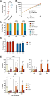
Figure 1.
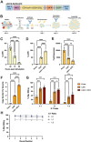
Figure 1.. Antibody-based stimulation yields robust outgrowth of polyclonal γδ T cell populations.
(A) Fold expansion after 11 days. Human peripheral blood γδ T cells were stimulated with either zoledronate or plate-bound αPan-γδ TCR and soluble αCD28 (n=11, paired T test). (B) Fold expansion at day 22 after two rounds of stimulation with αPan-γδ TCR and soluble αCD28 antibodies, occurring on day 0 and day 11 (n=11). (C) γδT cell subset frequency, as measured by flow cytometry, in zoledronate or αPan-γδ TCR plus soluble αCD28 stimulated γδ T cells and days 0, 11, and 22 (n=2 technical replicates of 2 independent biological donors). (D) Bar graphs showing Shannon entropy, a measure of TCR clonal diversity, of γδ T cell TCR diversity for TRD chains (red bars) and TRG chains (blue bars) at day 0, 11, and 22 (n=3). (E) Naive, TCM, TEM, and TEMRA frequencies in Vδ1+ (top left panel), Vδ2+ (top right panel), and Vδ1-Vδ2-αβ- (bottom panel) γδ T cells subsets as determined by CD27 and CD45ra expression (n=4). (****p<0.0001, **p<0.01, and *p<0.05)
Figure 2.
Figure 2.. Non-virally engineered CAR-γδ T cells display potent in vitro cytotoxicity.
(A) Diagram of construct containing CD19-CAR, mutant DHFR, conferring resistance to MTX in engineered cells, and GFP, used to manufacture CD19-CAR γδ T cells. (B) Diagram of our clinically scalable platform for γδ T cell engineering and expansion. Human γδ T cells are isolated by immunomagnetic separation from healthy donor PBMCs, then stimulated with plate-bound αPan-γδ TCR and soluble αCD28. γδ T cells are then electroporated with genome engineering reagents at day 2, re-stimulated on day 11, and collected at day 22. (C) Efficiency of non-viral construct delivery, as measured by GFP expression after stimulation and transfection of DNA-based CAR constructs alongside hyperactive Tc Buster transposase mRNA (n=8 for 48-hour time point, n=4 for 72- and 96-hour time points). (D) Frequency of CAR-γδ T cells, as measured by GFP expression, and (E) fold expansion on day 22 following MTX selection (n = 4). (F) CAR copy number per γδ T cell genome following non-viral integration as measured by ddPCR. Each point represents the average of three technical replicates (n=3). (G) In vitro cytolysis of Raji-luc cells after coculture with CD19-CAR γδ T cells. Assays were performed at 3:1, 1:1, and 1:3 E:T ratios in technical triplicate for two human donors. Luminescence intensity was used to quantify cytolysis of Raji-Luc target cells, with Triton X-100 serving as a positive control for max killing (n=3). (H) Cytotoxicity of CD19-CAR γδ T cells after repeated rounds of exposure to Raji-Luc target cells (n=6) (****p<0.0001, ***p<0.001 **p<0.01, and *p<0.05)
Figure 3.
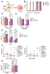
Figure 3.. Multiplex gene knockout enhances γδ T cell function in immunosuppressive environments.
(A) Diagram highlighting the roles of CISH, PDCD1, and FAS in suppressing γδ T cell activity in vivo. (B) Editing efficiency of CISH KO gRNA, PDCD1 KO gRNA, dnFAS gRNA, a combination of CISH and PDCD1 gRNA, and all three gRNAs, as well as a no-gRNA control in γδ T cells when cotransfected with CBE (n=4). Normalized survival of (C) unstimulated and (D) αPan-γδ TCR antibody-stimulated control, dnFAS, and triple edit CAR-γδ T cells either untreated or treated with soluble Fas ligand. Live cell counts were quantified after 24 hours and normalized to untreated control CAR γδ T cells (n=2). (E) Cytolysis of PDL1high Raji-Luc target cells following coculture with MTX-selected CAR γδ T cells, MTX-selected CAR γδ T cells with a PD1 KO, and MTX-selected CAR γδ T cells with PD1 KO, CISH KO, and Fas dn (n=6). (F) Fold expansion of engineered γδ T cells cultured in in decreasing amount of IL2 (left panel) or IL15 (right panel) (n=2). (G) Expression of 4–1BB, as measured by flow cytometry, in gene edited γδ T cells following stimulation with αPan-γδ TCR and soluble αCD28 for 24 hours (n=3).
Figure 4.
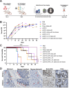
Figure 4.. Engineered polyclonal γδ T cells exhibit potent anti-tumor activity in vivo.
(A) Diagram of in vivo tumor challenge model. Mice were injected intravenously with 5×10⁶ Raji-luc cells on day −4 and treated with 10×106 γδ T cells on day 0. Mice that received two doses were injected again with 10×106 γδ T cells on day seven. Tumor growth and overall survival was measured and flow cytometry on blood was done weekly until they reached endpoint. (B) Tumor growth and (C) Kaplan-Meier survival curve for NSG mice injected with Raji-Luc cells and treated with the indicated γδ T cells (n=5). (D) Immunohistochemistry staining of CD3 on paraffin embedded skin, intestines, stomach, brain, and ovary in two animals treated with γδ T cells engineered with CD19-CAR and IL15.
Figure 5.
Figure 5.. Effect of gene engineering on γδ T cell subset frequency and persistence in vivo.
Weekly frequency of Vδ1+, Vδ2+, and Vδ1-Vδ2-αβ- subsets (A) as a percent of total CD3+ cells and (B) as a percent of total PBMC as measured by flow cytometry. “Infusion product” indicates the frequency before injection while “Endpoint” indicates the frequency at necropsy (n=10). (C) Frequency of γδ T cell subsets as a percent of total CD3+ cells and (D) as a percent of total PBMCs at necropsy as measured by flow cytometry (n=5). (E) Frequency of γδ T cell subsets as a percent of total CD3+ cells and (F) as a percent of total PBMCs at endpoint in groups that received a single dose of cells engineered with a constructs that did not contain IL15 compared with groups that received a single dose of cells engineered with constructs that contained IL15 (n=10).

References

    1. Davis M. M. & Bjorkman P. J. T-cell antigen receptor genes and T-cell recognition. Nature 334, 395–402 (1988). - PubMed
    1. Constant P. et al. Stimulation of human gamma delta T cells by nonpeptidic mycobacterial ligands. Science 264, 267–270 (1994). - PubMed
    1. Tanaka Y. et al. Natural and synthetic non-peptide antigens recognized by human gamma delta T cells. Nature 375, 155–158 (1995). - PubMed
    1. Fournié J. J. & Bonneville M. Stimulation of gamma delta T cells by phosphoantigens. Res. Immunol. 147, 338–347 (1996). - PubMed
    1. Gober H.-J. et al. Human T cell receptor gammadelta cells recognize endogenous mevalonate metabolites in tumor cells. J. Exp. Med. 197, 163–168 (2003). - PMC - PubMed

Publication types