Review
. 2024 Oct 25;5(11):e772.
doi: 10.1002/mco2.772. eCollection 2024 Nov.

Cardiomyopathy: pathogenesis and therapeutic interventions

Affiliations
Review

Cardiomyopathy: pathogenesis and therapeutic interventions

Shitong Huang et al. MedComm (2020). .

Abstract

Cardiomyopathy is a group of disease characterized by structural and functional damage to the myocardium. The etiologies of cardiomyopathies are diverse, spanning from genetic mutations impacting fundamental myocardial functions to systemic disorders that result in widespread cardiac damage. Many specific gene mutations cause primary cardiomyopathy. Environmental factors and metabolic disorders may also lead to the occurrence of cardiomyopathy. This review provides an in-depth analysis of the current understanding of the pathogenesis of various cardiomyopathies, highlighting the molecular and cellular mechanisms that contribute to their development and progression. The current therapeutic interventions for cardiomyopathies range from pharmacological interventions to mechanical support and heart transplantation. Gene therapy and cell therapy, propelled by ongoing advancements in overarching strategies and methodologies, has also emerged as a pivotal clinical intervention for a variety of diseases. The increasing number of causal gene of cardiomyopathies have been identified in recent studies. Therefore, gene therapy targeting causal genes holds promise in offering therapeutic advantages to individuals diagnosed with cardiomyopathies. Acting as a more precise approach to gene therapy, they are gradually emerging as a substitute for traditional gene therapy. This article reviews pathogenesis and therapeutic interventions for different cardiomyopathies.

Keywords: cardiomyopathy; disease‐causing gene; gene therapy; pathogenesis; personalized medicine; therapeutic interventions.


Conflict of interest statement

The authors declare no conflict of interest.

Figures
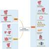
FIGURE 1
FIGURE 1
Classification of primary cardiomyopathies according to the AHA 2006 classification system. Primary cardiomyopathies can be divided into three categories: genetic, mixed, and acquired. Mixed cardiomyopathies include DCM and RCM. Acquired cardiomyopathy is the disease due to inflammatory stimulation and other factors, which mainly include four cardiomyopathies. Notably, recent evidence has emerged suggesting that a significant number of peripartum cardiomyopathy cases have genetic foundations. Genetic cardiomyopathy encompasses a collection of diseases resulting from gene mutations that induce abnormalities in both the structure and function of the cardiac muscles. There are many different types of genetic cardiomyopathies.
FIGURE 2
FIGURE 2
Mechanisms of general strategies and techniques in therapy of cardiomyopathy. (A) Viral vectors. In vivo, the viral vectors are delivered into the cardiac via IM injection and IC perfusion. In ex vivo, cells are extracted from the blood vessels of patients and seeded in a culture dish; after the viral capsid is removed, viruses are transduced in the culture containing patient cells, and they expand in the culture dish together. (B) iPSCs. iPSC cells can be modified with iPSC reprogramming factors or genome editing and differentiated into cardiomyocytes to be transplanted back into the patients. The iPSC‐derived cells can secrete exosomes, which mediate intercellular communication in heart exosomes. (C) CRISPR/Cas9 system. The Cas9 enzyme is used to cut two strands of DNA on a specific area as a pair of molecular scissors to remove or add DNA. gRNA can specifically identify the piece of DNA that needs to be cut. After the double‐strand DNA is broken, the broken DNA can be joined through NHEJ or HDR. (D) TALENs. The TALEN is composed of TALE protein, which contains customizable DNA‐binding domains (DBD) and nuclease domains of FokI dimerizes. Residues 12 and 13 (repeat variable diresidues (RVDs)) in TALE are responsible for the recognition of a specific base. The FokI nuclease bonds together to the protein through a wild‐type TALE sequence. TALEN‐ELD and TALEN‐KKR for heterodimeric TALENs include mutated Fok I dimerizes named ELD and KKR. ΔN and ΔC represent truncated N‐terminal and C‐terminal domains of TALEN. Left DBD and right DBD were customized to bind closely. All of the TALENs contain an SV40 nuclear localization signal (NLS).
FIGURE 3
FIGURE 3
Summary of genes associated with HCM and their function. The sarcomere is the most basic contractile unit in cardiac myocytes. Mutations in genes encoding sarcomeric proteins give rise to cardiomyopathies. The motor domains of myosin filaments (encoded by MYH6, etc.) cyclic interact with actin filaments (encoded by ACTN1, etc.) and form cross‐bridges, generating force and movement by using ATP, which influenced by intracellular Ca2+ concentration, PLN, and so on can regulate Ca2+ influx. Cardiac troponin is the protein that plays a major regulatory role in the contractile machinery. There are three subunits: troponin T (cTnT), binding to tropomyosin (encoded by TPM1), troponin I (cTnI), regulating actin ATPase activity, and troponin C (cTnC), binding to calcium, which separately encoded by TNNT2, and so on. The shortening of the sarcomere is caused by a relative sliding of the actin and myosin filaments. Cross‐linking elements such as Z‐disc (encoded by CSPR3, etc.) at the Z‐ and M‐lines hold the sarcomeric structures in the correct place. Titin proteins (encoded by TTN) connect the myosin filaments to the Z line leading to muscle relaxation and flexing. Cardiac myosin‐binding protein C, encoded by MYBPC3, is an important regulator of cardiomyocyte contraction and relaxation.
FIGURE 4
FIGURE 4
The causal genes in primary cardiomyopathies. (A) The shared genes between DCM and other cardiomyopathies. (B) The different mutations associated with RCM in patients, in vitro, and in animals. The different mutations have been found in patients, in vitro, and in animals. In different species, recent studies have found that mutations at different sites of the same gene can lead to RCM.

References

    1. Lipshultz SE, Law YM, Asante‐Korang A, et al. Cardiomyopathy in Children: Classification and Diagnosis: A Scientific Statement From the American Heart Association. Circulation. 2019;140(1):e9‐e68. - PubMed
    1. Dadson K, Hauck L, Billia F. Molecular mechanisms in cardiomyopathy. Clin Sci (Lond). 2017;131(13):1375‐1392. - PubMed
    1. Maron BJ, Towbin JA, Thiene G, et al. Contemporary definitions and classification of the cardiomyopathies: an American Heart Association Scientific Statement from the Council on Clinical Cardiology, Heart Failure and Transplantation Committee; Quality of Care and Outcomes Research and Functional Genomics and Translational Biology Interdisciplinary Working Groups; and Council on Epidemiology and Prevention. Circulation. 2006;113(14):1807‐1816. - PubMed
    1. Strong A, Musunuru K. Genome editing in cardiovascular diseases. Nat Rev Cardiol. 2017;14(1):11‐20. - PubMed
    1. Tan K, Foo R, Loh M. Cardiomyopathy in Asian Cohorts: Genetic and Epigenetic Insights. Circ Genom Precis Med. 2023;16(5):496‐506. - PubMed

LinkOut - more resources