. 2024 Oct 24;5(11):e742.
doi: 10.1002/mco2.742. eCollection 2024 Nov.

Partial reduction of interleukin-33 signaling improves senescence and renal injury in diabetic nephropathy

Affiliations

Partial reduction of interleukin-33 signaling improves senescence and renal injury in diabetic nephropathy

Li Chen et al. MedComm (2020). .

Abstract

Diabetic nephropathy (DN) is a frequent and costly complication of diabetes with limited understandings of mechanisms and therapies. Emerging evidence points to the important roles of interleukin-33 (IL-33) in acute kidney injury, yet its contribution to DN is still unclear. We here found a ubiquitous increase of IL-33 and its receptor (ST2) in murine models and patients with DN. Surprisingly, both IL-33 and ST2 knockdown aggravated renal lesions in DN, while overexpression of IL-33 also exacerbated the condition. Further population-based analyses revealed a positive correlation of IL-33 expression with renal dysfunction in DN patients. Individuals with high IL-33 expression-related polygenic risk score had a higher DN risk. These findings confirmed the harmful effects of IL-33 on DN. Conversely, endogenous and exogenous partial reduction of IL-33 signaling conferred renoprotective effects in vivo and in vitro. Mechanistically, IL-33 induced senescence by regulating cell cycle factors in HK-2 cells, and accordingly senescence led to renal cell damage through the secretion of senescence-related secretory phenotype (SASP) including IL-33 and prostaglandins. Together, elevated IL-33 accelerates cellular senescence to drive DN possibly by SASP production, while a partial blockage improves renal injury and senescence. Our findings pinpoint a possible and new avenue for DN interventions.

Keywords: cellular senescence; diabetic nephropathy; interleukin‐33; senescence‐related secretory phenotype.


Conflict of interest statement

All authors declared no conflict of interest.

Figures

FIGURE 1
FIGURE 1
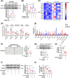
IL‐33 and ST2 levels are increased in human participants and animals with DN. (A) Western immunoblot analysis for IL‐33 and ST2 in kidney of DN mice (HFD+STZ), ZDF rats, or ApoE−/− mice fed with HFD and respective control animals. GAPDH served as a loading control. The third blot shows the shorter IL‐33. (B and C) Serum IL‐33 levels and the expression levels of IL‐33 and ST2 mRNA in DN mice (HFD+STZ) and control mice (ND). (D) The protein expression of IL‐33 and ST2 in the paracancerous kidneys and the kidneys of DN patients was shown by immunohistochemistry (scale bar: 20 µm). (E) Fold change of IL‐33 mRNA levels in DN‐related models compared with control kidney tissue. Data from the GEO database. (F) At 2, 4, and 8 months, renal mRNA levels of IL‐33 in OVE26 diabetic mice and its control group (n = 4–7). (G) At 10 and 20 weeks, IL‐33 mRNA levels in the kidney of STZ‐induced diabetic rats and its control group (n = 3). (H) Renal IL‐33 mRNA expression in non‐diabetic, early‐stage, and advanced DN individuals. *p < 0.05, **p < 0.01, ***p < 0.001.
FIGURE 2
FIGURE 2
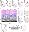
Renal injury is exacerbated in IL‐33−/− or ST2−/− DN mice. WT, IL‐33−/−, and ST2−/− mice were continued to be fed for 12 weeks after they were determined to be diabetic. Control mice were continuously fed with ND. (A–E) FBG, renal‐body ratio, Scr, BUN, and ACR were monitored at the end of the experiment. (F) Representative images of kidney tissue stained with H&E and PAS (scale bar: 20 µm). Ultra‐structural changes in glomerular morphology were assessed by transmission electron microscopy (scale bar: 1 µm). (G and H) The glomerular area and PAS‐positive area were assessed in 3 mice with at least 10 glomeruli per mouse. (I and J) GBM thickness and foot process width were calculated in electron microscopic images of at least 5 fields of view per mouse. (K) The renal tubular injury score was estimated by H&E staining. (L) Immunoblot analysis for KIM‐1. *p < 0.05, **< 0.01, ***p < 0.001.
FIGURE 3
FIGURE 3
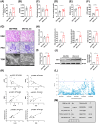
rIL‐33 administration aggravates renal injury in DN mice. Diabetic mice were intraperitoneally injected with PBS or rIL‐33 twice a week for 12 weeks and maintained with HFD feeding. (A) Serum IL‐33 levels were measured by ELISA at the endpoint of the experiment. (B–F) FBG, renal‐body ratio, Scr, BUN, and ACR were assessed. (G) Representative micrographs about DN‐related pathological indicators. (H) The statistics of glomerular area, PAS‐positive area, GBM thickness, and foot process width. (I) Renal tubular injury score based on the area of renal tubular injury in H&E images. (J) Renal lysates were processed for western immunoblot analysis for KIM‐1. (K) Correlation of eGFR, glomerular area, and KIM‐1 expression with IL‐33 level and ST2 level. (L) eQTL analysis between IL‐33 mRNA in whole blood and the SNPs of IL33 nearby region (± 500 kb) using data provided by GTEx. The x‐axis showed the chromosomal positions and the y‐axis showed a −log10 p value; the significance threshold used in our analysis was < 0.01. (M) OR (95% CI) in PRS associated with DN risk among participants with or without DN from UK biobank. *p < 0.05, **p < 0.01, ***p < 0.001.
FIGURE 4
FIGURE 4
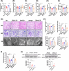
Renal senescence and aging are improved in IL‐33+/− and ST2+/− DN mice and. αIL‐33‐treated WT DN mice. WT mice considered diabetic were intraperitoneally injected with IgG or αIL‐33 twice a week for 12 weeks and maintained with HFD feeding. IL‐33+/− and ST2+/− DN mice were maintained with HFD feeding for 26 weeks. (A) The circulating IL‐33 level was measured by ELISA. (B and C) FBG and renal‐body ratio. (D‐F) Renal function was measured by Scr, BUN, and ACR. (G) Representative images of H&E, PAS staining (scale bar: 20 µm), and transmission electron microscopy (scale bar: 1 µm). (H) The statistics of pathological indicators in the glomeruli included glomerular area, PAS‐positive areas, GBM thickness, and foot process width. (I and J) Renal tubular injury score and KIM‐1 protein level were used to measure renal tubular changes. *< 0.05, **p < 0.01, ***p < 0.001.
FIGURE 5
FIGURE 5
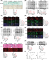
rIL‐33 exacerbates renal senescence and aging in DN mice. (A) Representative images of kidney tissue stained with SA‐β‐gal and Ki67. The statistical data were the ratio of positive area. (B) The protein levels of senescence markers were determined by immunoblots. Densitometric analysis of p53, p21, p16, ATM, and γH2AX. (C and D) Representative fluorescence colocalization images of IL‐6 (red) and NF‐κB (red) with SA‐β‐gal (C12FDG, green) in renal tissue. Colocalization statistics were shown as Pearson's correlation coefficient. (E) Protein levels in the cell apoptosis pathway, including AKT, p‐AKT, Bcl‐2, Bax, and Caspase‐3. (F) Colocalization of the prosurvival protein Bcl‐2 (red) with SA‐β‐gal (green). (G) The extent of fibrosis (Masson staining) and the level of oxidative stress in the kidneys (ROS fluorescent probe, 10 µM). (H) Fibrosis‐related protein levels including Collagen I and CTGF. (I) Correlation of p16 expression with IL‐33 and ST2 level in DN patients. Scale bar: 20 µm. *p < 0.05, **p < 0.01, ***p < 0.001.
FIGURE 6
FIGURE 6
Partial reduction of IL‐33 alleviates cell injury and senescence, and cellular senescence mediates the role of IL‐33 in vitro. (A and B) KIM‐1 expression and LDH release in cells treated with different doses of αIL‐33 (10 or 50 ng/mL) and rIL‐33 (10 or 50 ng/mL) for 10 days. (C and D) cellular senescence‐related changes including the ratio of SA‐β‐gal+ and EdU+ cells, and fold change in nuclear area and γH2AX expression. (E) The protein levels of p53, p21, and p16 in HK‐2 cells. (F–H) Cell damage‐related (KIM‐1 expression and LDH release) and cellular senescence‐related changes (SA‐β‐gal, EdU, nuclear area, and γH2AX) in rIL33‐treated (50 ng/mL) cells with p16 and p21 silencing respectively or simultaneously. (I–K) SA‐β‐gal, Hochest (marked apoptotic cells), and LDH analyses of HK‐2 cells exposed to HG for 17 days and ABT‐263 (10 µM) for 1 day. Scale bar: 20 µm. *p < 0.05, **p < 0.01, ***p < 0.001.
FIGURE 7
FIGURE 7
SASP derived from senescent cells contributes to renal cell damage. (A and B) KIM‐1 expression and LDH release in young HK‐2 cells treated with different CM‐SASP for 2 days. (C and D) Prostaglandins levels in DN mice with rIL‐33 or αIL‐33. (E) Prostaglandin levels in SASP of senescent HK‐2 cells (n = 3). (F–H) SA‐β‐gal+ cell, KIM‐1 expression, and LDH release in HK‐2 cells treated with COX‐2 inhibitor NS‐398 (10 µM) for 5 days (scale bar: 20 µm). (I and J) KIM‐1 expression and LDH release in young HK‐2 cells treated with different CM‐SASP (Si‐IL‐33 and NS‐398) for 2 days. *p < 0.05, **p < 0.01, ***p < 0.001.
FIGURE 8
FIGURE 8
Mechanism flow chart. Sustained hyperglycemic stimulation leads to an increased production of IL‐33, which in turn accelerates senescence in tubular epithelial cells. The senescent cells then release SASP components, including IL‐33 and prostaglandins, which further amplify cellular senescence and exacerbate renal lesions.

References

    1. Cao R, Tian H, Zhang Y, et al. Signaling pathways and intervention for therapy of type 2 diabetes mellitus. MedComm. 2023;4(3):e283. - PMC - PubMed
    1. Tuttle KR, Agarwal R, Alpers CE, et al. Molecular mechanisms and therapeutic targets for diabetic kidney disease. Kidney Int. 2022;102(2):248‐260. - PubMed
    1. DeFronzo RA, Reeves WB, Awad AS. Pathophysiology of diabetic kidney disease: impact of SGLT2 inhibitors. Nat Rev Nephrol. 2021;17(5):319‐334. - PubMed
    1. Cayrol C, Girard JP. Interleukin‐33 (IL‐33): a nuclear cytokine from the IL‐1 family. Immunol Rev. 2018;281(1):154‐168. - PubMed
    1. Li T, Zhang Z, Bartolacci JG, et al. Graft IL‐33 regulates infiltrating macrophages to protect against chronic rejection. J Clin Invest. 2020;130(10):5397‐5412. - PMC - PubMed

LinkOut - more resources