. 2024 Oct 10;1(4):ugae015.
doi: 10.1093/narmme/ugae015. eCollection 2024 Oct.

C G composition in transposon-derived genes is increased in FXD with perturbed immune system

Affiliations

C G composition in transposon-derived genes is increased in FXD with perturbed immune system

Tamaki Suganuma et al. NAR Mol Med. .

Abstract

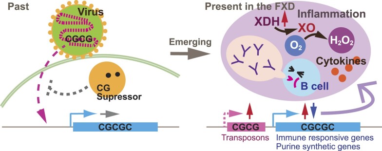
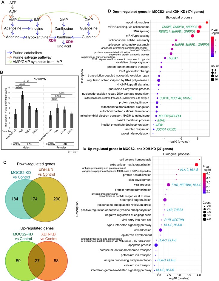
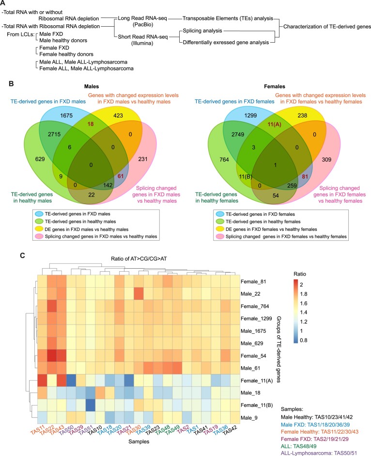
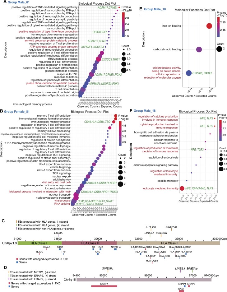
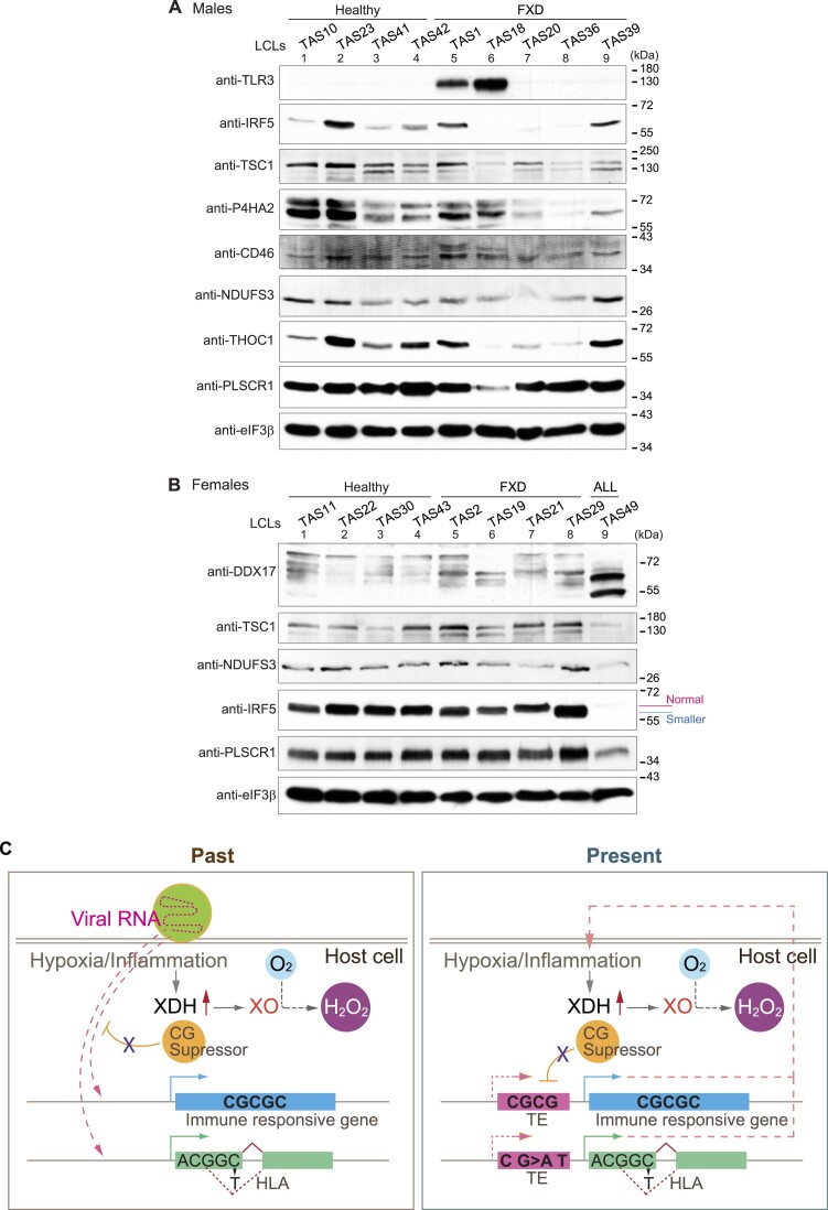
Increasing incidence of Fragile X disorders (FXD) and of immune-mediated disorders in FXD suggests that additional factors besides FMR1 mutations contribute to the pathogenesis. Here, we discovered that the expression levels or splicing of specific transposon element (TE)-derived genes, regulating purine metabolism and immune responses against viral infections are altered in FXD. These genes include HLA genes clustered in chr6p21.3 and viral responsive genes in chr5q15. Remarkably, these TE-derived genes contain a low A T/C G suggesting base substitutions of A T to C G. The TE-derived genes with changed expression levels contained a higher content of 5'-CG-3' dinucleotides in FXD compared to healthy donors. This resembles the genomes of some RNA viruses, which maintain high contents of CG dinucleotides to sustain their latent infection exploiting antiviral responses. Thus, past viral infections may have persisted as TEs, provoking immune-mediated disorders in FXD.


Figures

Graphical Abstract
Graphical Abstract
Figure 1.
Figure 1.
XDH and MOCS2 are essential for translations of genes regulating responses to viral infection and oxidative phosphorylation. (A) Purine catabolism. (B) Xanthine oxidase (XO) activity in cytoplasmic extracts of LCLs were measured by fluorometric assay (mean ± SD, n = 2). Averaged XO activities in healthy donor cells were compared with each FXD cells in each gender. The numbers of two-tailed test (T.TEST) are indicated. (C) Area-proportional venn diagrams indicating the numbers of genes with significantly changed transcript levels (adjusted P-value <0.05 and fold change >2) in polysomes in MOCS2- or XDH-knockdown cells compared to controls (n = 2 or 3). (D and E) Dot plots display GO analysis clustered by biological process of genes with decreased (D) or increased (E) transcripts in polysomes in MOCS2- and XDH-knockdown cells compared to polysomes in controls by RNA-seq. Representative genes are indicated.
Figure 2.
Figure 2.
Low A T / C G leads to altered expressions of TE-derived genes in FXD. (A) Diagram of the RNA-seq strategy used in this study. (B) Venn diagram shows transposable elements (TEs)-derived genes identified using PacBio-reads and overlapped with splicing changed genes and genes with changed expression levels (differentially expressed (DE) genes) identified using Illumina-short reads, in males (left) and females (right). (A) and (B) are indicated to distinguish between each group of 11 genes for convenience. (C) Heatmap with rows for each group of genes corresponding to those in (B) and columns for samples displays the raw data of ratio of AT > CG / CG > AT, which were analyzed by the base substitutions of each nucleotide (A T > C G: sum of A > C, A > G, T > C and T > G base substitutions; C G > A T: sum of C > A, C > T, G > A and G > T base substitutions), in averaged sum (n = 3) of the base substitutions.
Figure 3.
Figure 3.
5′-CG-3′ are increased in TE-derived genes with altered expressions in FXD. (AB) Box plots display the count of CG di-nucleotides normalized to length of the gene in 1000 bp using the ATGC consensus in each group of TE-derived genes corresponding to the groups of Venn diagram in Figure 2B (Supplementary Figures S3 and S4). Sample names and repeats are indicated at the bottom. The count in each repeat (n = 3) is indicated. ALL*: ALL-lymphosarcoma.
Figure 4.
Figure 4.
Expressions of TE-derived genes regulating purine metabolism and immune responses were changed in FXD. (A, B, E, F) Dot plots display GO analysis clustered by biological process (A, B, F) or molecular functions (E) for TE-derived genes with changed splicing corresponding to group male 61 (A) or group female 81 (B) in Figure 2B (P < 0.05) or changed transcript levels corresponding to group male 18 (E and F) in Figure 2B (adjusted P < 0.05) by using RNA-seq. The TOP 30 processes are listed in A and B. Representative genes are indicated. (C, D) Locations of HLA genes (C) and ERAP2 (D) with/without changed expressions and TEs (Alu, LTR, LINE, and SINE) are illustrated.
Figure 5.
Figure 5.
Protein expressions of TE-derived genes responding viral infection were changed in FXD. (A, B) Protein expressions of TE-derived genes regulating immune responses (TLR3, IRF5, TSC1, CD46 and THOC1), viral responses (PLSCR1 and DXX17), oxidoreductase activity (P4HA2), and mitochondrial function and purine metabolism (NDUFS3) in the extracts from indicated LCLs were analyzed by western blots. eIF3β was used as the loading control. (C) Summary model is illustrated. Viral RNA with a high content of CG dinucleotides is integrated into the host genome by exploiting CG suppressor to decrease antigens and is retained as retrotransposons (TEs) (left panel). Some viral infections trigger hypoxia, which promotes conversion of O2 to H2O2 via XO (left panel). Inflammation induces XDH expression and conversion of XDH to XO, producing ROS including H2O2 (left and right panels). In FXD in the present, increased substitution of A T to C G in TE-derived genes causes splicing changes, including in HLA genes (right panel). Increased CG dinucleotide changes expression of TE-derived genes regulating immune responses, suggesting that these genes had deceived CG suppression. Thus, maintaining high levels of C G content and CG dinucleotides in TE-derived genes likely stimulates XO activity and has maintained expression of TEs.

References

    1. Sokol D.K., Maloney B., Long J.M., Ray B., Lahiri D.K. Autism, Alzheimer disease, and fragile X: APP, FMRP, and mGluR5 are molecular links. Neurology. 2011; 76:1344–1352. - PMC - PubMed
    1. Hagerman P.J., Hagerman R.J. Fragile X-associated tremor/ataxia syndrome. Ann. N. Y. Acad. Sci. 2015; 1338:58–70. - PMC - PubMed
    1. Herring J., Johnson K., Richstein J. The use of “retardation” in FRAXA, FMRP, FMR1 and other designations. Cells. 2022; 11:1044–1050. - PMC - PubMed
    1. Wheeler A.C., Bailey D.B. Jr., Berry-Kravis E., Greenberg J., Losh M., Mailick M., Mila M., Olichney J.M., Rodriguez-Revenga L., Sherman S et al. . Associated features in females with an FMR1 premutation. J. Neurodev. Disord. 2014; 6:30. - PMC - PubMed
    1. Yu K.H., Palmer N., Fox K., Prock L., Mandl K.D., Kohane I.S., Prilutsky D. The phenotypical implications of immune dysregulation in fragile X syndrome. Eur. J. Neurol. 2020; 27:590–593. - PubMed

LinkOut - more resources