. 2024 Oct 27;14(1):25682.
doi: 10.1038/s41598-024-76258-4.

Targeting the VEGFR2 signaling pathway for angiogenesis and fibrosis regulation in neovascular age-related macular degeneration

Affiliations

Targeting the VEGFR2 signaling pathway for angiogenesis and fibrosis regulation in neovascular age-related macular degeneration

Eunhye Yu et al. Sci Rep. .

Abstract

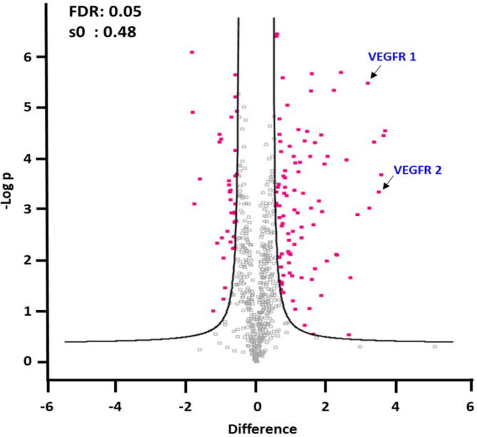
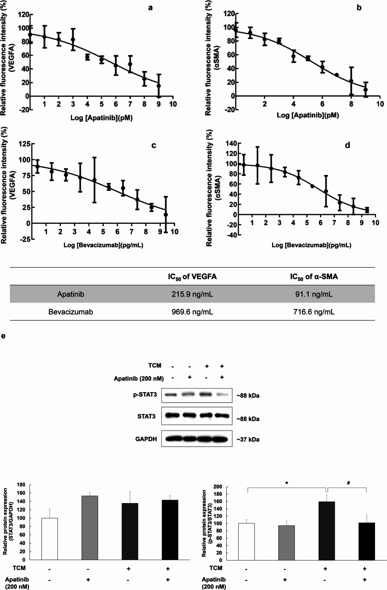
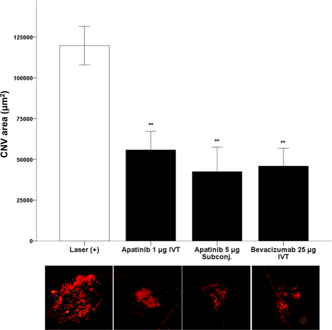
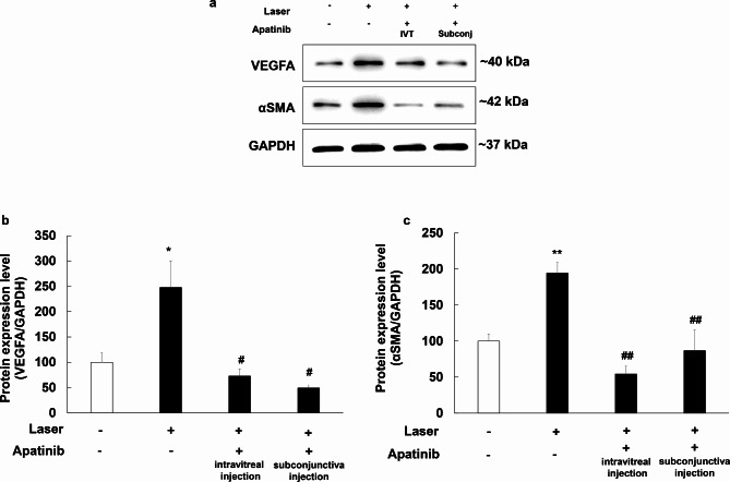
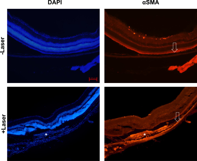
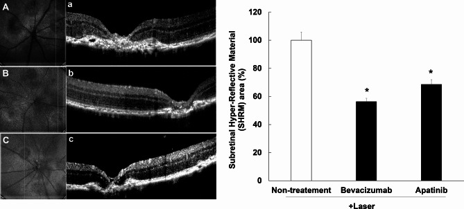
Neovascular age-related macular degeneration (nAMD) is characterized by abnormal blood vessel growth from the choroid, leading to complications and eventual blindness. Despite anti-VEGF therapy, subretinal fibrosis remains a major concern, as VEGF/VEGF receptor-2 (VEGFR2) signaling can contribute to both angiogenesis and fibrosis. For the identification of the aqueous humor proteome, we performed liquid chromatography with tandem mass spectrometry analysis. To investigate the potential therapeutic effects of targeting the VEGF signaling pathway using apatinib, a highly selective VEGFR2 tyrosine kinase inhibitor, this study employed in vitro (THP-1 conditioned media-treated ARPE-19 cells) and in vivo (laser-induced choroidal neovascularization mouse) models of nAMD. This study revealed elevated VEGFR2 protein levels in the aqueous humor of nAMD patients, suggesting a potential target to mitigate neovascularization and fibrosis in nAMD. Apatinib effectively reduced VEGFA and αSMA levels in both in vitro and in vivo models. Moreover, apatinib showed improvement in laser-induced subretinal hyper-reflective lesions. The action mechanism was linked to the inhibition of VEGFR2 activation, leading to the suppression of both angiogenesis and fibrosis through the downregulation of STAT3 phosphorylation. Therefore, the VEGFR2 signaling pathway appears to play a central role in the development of nAMD by regulating both angiogenesis and fibrosis.

Keywords: Apatinib; Choroidal neovascularization; Fibrosis; Neovascular age-related macular degeneration; Tyrosine kinase inhibitor; Vascular endothelial growth factor receptor-2.


Conflict of interest statement

The authors declare no competing interests.

Figures

Fig. 1
Fig. 1
Apatinib, a small-molecule receptor tyrosine kinase inhibitor, exhibits potential antiangiogenic and antineoplastic effects. It selectively binds to and inhibits vascular endothelial growth factor (VEGF) receptor 2, thereby potentially suppressing VEGF-stimulated endothelial cell migration and proliferation, consequently reducing neovascularization.
Fig. 2
Fig. 2
Differentially expressed proteins in the aqueous humor of patients with nAMD and control group. Quantitation of VEGFR1 and VEFGR2, which trigger the angiogenic action of VEGF, showed that they significantly upregulated in nAMD compared to control. The cutoff criteria for protein quantitation were as follows: t-test, p < 0.05, minimum confidence level > 95%, more than two unique peptides, FDR < 5%, and fold-change thresholds (expressed as log2 ratio) of > 1.5 or < − 1.5. For the analysis of differentially expressed proteins and statistical analysis, Perseus (version 1.5.8.2) was used, where the cutoff criteria for significant fold change (FC) and t-test p values were set at ± 1.5 and 0.05, respectively.
Fig. 3
Fig. 3
The half maximal inhibitory concentration (IC50) values of apatinib against VEGFA (a) and αSMA (b), bevacizumab against VEGFA (c) and αSMA (d). For afatinib, the IC50 values were 215.9 ng/mL for VEGFA and 91.1 ng/mL for αSMA. For bevacizumab, the IC50 values were 969.6 ng/mL for VEGFA and 716.6 ng/mL for αSMA. The data were fitted to log (concentration) and normalized response equations. The results are expressed as mean ± S.E. (n = 3). Suppressive effects of apatinib on THP-1-conditioned media (TCM)-induced STAT3 phosphorylation in ARPE-19 cells (e). Western blot analysis was used to examine the expression of p-STAT3 in ARPE-19 cells. ARPE-19 cells were stimulated by TCM for 3 h in 6-well plates and then treated with apatinib (200 nM) for 24 h. Thereafter, the whole lysate was analyzed for protein expression. Data were analyzed using two-way ANOVA (*p < 0.05 vs. untreated group, #p < 0.05 vs. only TCM (+) group), and the results are expressed as mean ± S.E. (n = 3). Uncropped blot images are presented in supplementary information (Supplementary Fig. S3).
Fig. 4
Fig. 4
Change in the laser-induced choroidal neovascularization (CNV) area after intravitreal treatment (IVT) or subconjunctival treatment (subconj.) with apatinib and bevacizumab in mice with laser-induced CNV. Laser setting (300 mW, 100 ms, 95% white bubble, 78 D lens). CNV was stained using Alexa Fluor 594-conjugated isolectin B4 (1:100 dilution). Data were analyzed using the Mann–Whitney U test (**p < 0.01 vs. only laser-induced CNV) and are expressed as the mean ± S.E. (n = 6–17).
Fig. 5
Fig. 5
Inhibitory effects of apatinib on VEGFA (b) and αSMA (c) expression in the mouse model of laser-induced choroidal neovascularization (CNV). Intravitreal (IVT) injection of apatinib (1 µg in 1 µL DMSO) and subconjunctival (subconj) injection of apatinib (5 µg in 1 µL DMSO) were administered immediately after laser photocoagulation. Equal amounts of protein from eye tissue lysates of mice with laser-induced CNV were analyzed for the expression of the indicated proteins. Protein expression in the eyes of mice with laser-induced CNV was measured 1 week after laser photocoagulation by Western blot analysis (a). Data were analyzed using one-way ANOVA (*p < 0.05 and **p < 0.01 vs. non-laser exposed group, #p < 0.05 and ##p < 0.01 vs. only laser exposed group). The results are expressed as mean ± S.E. (n = 5–12). Uncropped blot images are presented in supplementary information (Supplementary Fig. S4).
Fig. 6
Fig. 6
Expression of αSMA in the eyes of mice with laser-induced choroidal neovascularization (CNV). Immunohistochemical staining of the eyes of mice with and without laser-induced CNV (DAPI, blue color and αSMA, orange color). White asterisks indicate subretinal fibrosis and deposits and increased αSMA expression level. The white arrow depicts the retinal pigment epithelium layer. Scale bar for each image: 100 μm.
Fig. 7
Fig. 7
Inhibitory effects of apatinib on the expression of hyper-reflective lesions in mice with laser-induced choroidal neovascularization (CNV). Laser photocoagulation spots were identified in digital red-free retinal images (non-treatment: (A) bevacizumab: (B) apatinib: (C). Using the center of the laser spot as reference, retinal OCT scans were performed in the vertical plane (non-treatment: (a) bevacizumab: (b) apatinib: (c). Hyper-reflective lesions were confirmed on OCT images. Thereafter, the inner border of the lesion was manually outlined, whereas the hyper-reflective area (mean area of vertical and horizontal planes) was measured using Image J software (version 1.53, https://imagej.nih.gov/ij/index.html). The hyper-reflective lesion area was significantly decreased after intravitreal administration of apatinib (1 µg/1 µL in DMSO) and bevacizumab as positive control (25 µg/1 µL) (D). Data were analyzed using the Kruskal–Wallis test followed by Dunn’s test (*p < 0.05 vs. only laser-induced CNV group), and the results are expressed as mean ± S.E. (n = 10–29).

References

    1. Lambert, N. G. et al. Risk factors and biomarkers of age-related macular degeneration. Prog. Retin. Eye Res.54, 64–102. 10.1016/j.preteyeres.2016.04.003 (2016). - PMC - PubMed
    1. Witmer, A. N., Vrensen, G. F., Van Noorden, C. J. & Schlingemann, R. O. Vascular endothelial growth factors and angiogenesis in eye disease. Prog. Retin. Eye Res.22, 1–29. 10.1016/s1350-9462(02)00043-5 (2003). - PubMed
    1. Heier, J. S. et al. Ranibizumab for treatment of neovascular age-related macular degeneration: A phase I/II multicenter, controlled, multidose study. Ophthalmology113(633), e631-634. 10.1016/j.ophtha.2005.10.052 (2006). - PubMed
    1. Rosenfeld, P. J. et al. Ranibizumab for neovascular age-related macular degeneration. N. Engl. J. Med.355, 1419–1431. 10.1056/NEJMoa054481 (2006). - PubMed
    1. Heier, J. S. et al. Intravitreal aflibercept (VEGF trap-eye) in wet age-related macular degeneration. Ophthalmology119, 2537–2548. 10.1016/j.ophtha.2012.09.006 (2012). - PubMed

MeSH terms