. 2024 Oct 27;15(1):9269.
doi: 10.1038/s41467-024-53614-6.

Mechanistic basis for the allosteric activation of NADase activity in the Sir2-HerA antiphage defense system

Affiliations

Mechanistic basis for the allosteric activation of NADase activity in the Sir2-HerA antiphage defense system

Xiangkai Zhen et al. Nat Commun. .

Abstract

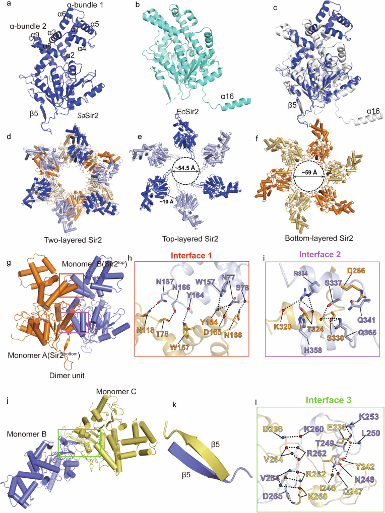
Sir2-HerA is a widely distributed antiphage system composed of a RecA-like ATPase (HerA) and an effector with potential NADase activity (Sir2). Sir2-HerA is believed to provide defense against phage infection in Sir2-dependent NAD+ depletion to arrest the growth of infected cells. However, the detailed mechanism underlying its antiphage activity remains largely unknown. Here, we report functional investigations of Sir2-HerA from Staphylococcus aureus (SaSir2-HerA), unveiling that the NADase function of SaSir2 can be allosterically activated by the binding of SaHerA, which then assembles into a supramolecular complex with NADase activity. By combining the cryo-EM structure of SaSir2-HerA in complex with the NAD+ cleavage product, it is surprisingly observed that Sir2 protomers that interact with HerA are in the activated state, which is due to the opening of the α15-helix covering the active site, allowing NAD+ to access the catalytic pocket for hydrolysis. In brief, our study provides a comprehensive view of an allosteric activation mechanism for Sir2 NADase activity in the Sir2-HerA immune system.


Conflict of interest statement

The authors declare no competing interests.

Figures

Fig. 1
Fig. 1. Sir2 alone in SaSir2-HerA does not exhibit NADase activity, which can be activated upon the binding of HerA.
a Schematic diagram of the Sir2 and HerA operon of S. aureus. b In vitro NADase activity of SaSir2 and SaSir2-HerA; SaSir2 could not hydrolyze NAD+; in contrast, a mixture of SaSir2 and SaHerA exhibited NADase activity. c The gel filtration profile and SDS‒PAGE results of copurified SaSir2-HerA. d AUC result for apo SaSir2, suggesting that it behaved as a homodimer in solution. e Effects of different concentrations of ATP on the NADase activity of SaSir2-HerA. Data were presented as mean ± SD in histograms; n = 3 independent experiments. P-value determined by paired two-tailed t-tests is indicated in the figure. Source data are provided as a Source Data file.
Fig. 2
Fig. 2. Cryo-EM structure of the homotrimeric SaHerA.
a Cryo-EM density maps of the trimeric SaHerA from different views. b Ribbon diagram of the structure of the homotrimeric SaHerA from the same views as in (a). c Structure of SaHerA generated by AlphaFold; the HAS domain, ATPase domain and helical bundle are colored orange, cyan and blue, respectively. d The portions discernible in the apo SaHerA structure are boxed. e, f Detailed interactions among the protomers mediated by the helical-bundle domain of SaHerA.
Fig. 3
Fig. 3. Cryo-EM structure of the SaSir2-HerA supramolecular complex.
a Different views of the cryo-EM density map of the SaSir2-HerA complex, SaSir2 and SaHerA are shown with the protomers color coded. b Atomic model built into the cryo-ME map, which is colored as (a). c, d Cartoon mode and surface mode of the N-terminal domain of SaHerA. e Top view of the SaHerA hexamer; the two clefts between the two trimers are illustrated.
Fig. 4
Fig. 4. Structural analysis of SaSir2 domain and the assembly of the two-layered SaSir2 dodecamer.
a SaSir2 exhibits a typical Rossmann fold. b The closest structurally similar SaSir2 NADase (EcSir2, PDB 8SXX) to SaSir2. c Overlay of SaSir2 and EcSir2 highlighting the differences among SaSir2, EcSir2 and other Sir2 domain-containing proteins. The additional loop and α helix bundle are highlighted in blue. d Cartoon model of the SaSir2 dodecamer. e The hexameric layer of SaSir2 protomers that interact directly with SaHerA (upper layer). f The bottom layer of SaSir2 protomers that do not interact with SaHerA. g Homodimer units of SaSir2. h-i Enlarged view of the interactions shown in (g). j Neighboring SaSir2 dimer unit were pulled together by Sir2 molecules in the bottom layer (Molecule B) and the top layer (Molecule C). k Enlarged view of the β-hairpin formed in (j). l Detailed interactions of the interfaces shown in (j).
Fig. 5
Fig. 5. Interactions between SaSir2 and SaHerA.
a Surface and cartoon representation of SaSir2-HerA, showing the minimal SaSir2-HerA unit. b, c Specific interactions between SaSir2 and SaHerA; the residues involved in their interactions are shown as sticks. d Deletion of the N-terminal residues of Sir2 and HerA, encompassing residues 1–34 and 1–35, simultaneously abolished the NADase activity of Sir2-HerA. Data were presented as mean ± SD in histograms from three replicates. P-values were determined by paired two-tailed t-tests. e In vitro pull-down assays of SaSir2 with an N-terminal 1–34 deletion and HerA with an N-terminal 1–35 deletion, His-tagged HerA was used as bait, Sir2 was used as a pray. Source data are provided as a Source Data file.
Fig. 6
Fig. 6. Mechanism of NAD+ catalysis of SaSir2 NADase.
a Cut-open sliced view of the NAD+ binding pocket of SaSir2top, the NAD+ cleavage product is shown in sticks. b An enlarged view of ADPR coordination by the key residues of SaSir2. c Effects of mutations on the ADPR-coordinated residues in the in vitro NADase assays. Data were presented as mean ± SD in histograms from three replicates. P-values were determined by paired two-tailed t-tests. Source data are provided as a Source Data file.
Fig. 7
Fig. 7. The allosteric activation mechanism of SaSir2 NADase activity.
a Overlay of SaSir2 in the bottom layer (ADPR-free) and the upper layer (ADPR-bound) in cartoon representation. The SaSir2 molecules in the bottom and upper layers are colored white and cyan, respectively. α15 (residues 316-325) is colored blue, and the corresponding loop is highlighted in red. b Structural alignment of SaSir2top docked into NAD+ and SaSir2bottom (left) and enlarged view of the steric clashes between the NAD+ and SaSir2bottom, the NAD+ is shown in dotted surface mode (right). c, d BLI sensorgrams of SaSir2, SaSir2-HerA and NAD+. From the top curve to the bottom curve, NAD+ was subjected to twofold serial dilutions starting at 16.5 µM. e In vitro NADase activity of wild-type Sir2 and Sir2 with α15 deletion, as similar to the complex of SaSir2-HerA, the NAD+ level in SaSir2 with α15 deletion is significant lower that the wild-type SaSir2. Data were presented as mean ± SD in histograms from three replicates. Unpaired two-tailed Student’s tests were performed. f The proposed mechanism of the antiphage function of SaSir2-HerA. Source data are provided as a Source Data file.

References

    1. Georjon, H. & Bernheim, A. The highly diverse antiphage defence systems of bacteria. Nat. Rev. Microbiol. 21, 686–700 (2023). - PubMed
    1. Watson, B. N. J., Steens, J. A., Staals, R. H. J., Westra, E. R. & van Houte, S. Coevolution between bacterial CRISPR-Cas systems and their bacteriophages. Cell Host Microbe29, 715–725 (2021). - PubMed
    1. Hille, F. et al. The biology of CRISPR-Cas: backward and forward. Cell172, 1239–1259 (2018). - PubMed
    1. Doron, S. et al. Systematic discovery of antiphage defense systems in the microbial pangenome. Science359, eaar4120 (2018). - PMC - PubMed
    1. Gao, L. et al. Diverse enzymatic activities mediate antiviral immunity in prokaryotes. Science369, 1077–1084 (2020). - PMC - PubMed

Publication types

LinkOut - more resources