Clinical Trial
. 2025 Feb 3;31(3):503-514.
doi: 10.1158/1078-0432.CCR-24-2172.

Phase I Study of ROR1-Specific CAR-T Cells in Advanced Hematopoietic and Epithelial Malignancies

Affiliations
Clinical Trial

Phase I Study of ROR1-Specific CAR-T Cells in Advanced Hematopoietic and Epithelial Malignancies

Carla A Jaeger-Ruckstuhl et al. Clin Cancer Res. .

Abstract

Purpose: The receptor tyrosine kinase-like orphan receptor 1 (ROR1) is expressed in hematopoietic and epithelial cancers but has limited expression on normal adult tissues. This phase I study evaluated the safety of targeting ROR1 with autologous T lymphocytes engineered to express a ROR1 chimeric antigen receptor (CAR). Secondary objectives evaluated the persistence, trafficking, and antitumor activity of CAR-T cells.

Patients and methods: Twenty-one patients with ROR1+ tumors received CAR-T cells at one of four dose levels: 3.3 × 10⁵, 1 × 10⁶, 3.3 × 10⁶, and 1 × 10⁷ cells/kg body weight, administered after lymphodepletion with cyclophosphamide/fludarabine or oxaliplatin/cyclophosphamide. Cohort A included patients with chronic lymphocytic leukemia (CLL, n = 3); cohort B included patients with triple-negative breast cancer (TNBC, n = 10) or non-small cell lung cancer (NSCLC, n = 8). A second infusion was administered to one patient in cohort A with residual CLL in the marrow and three patients in cohort B with stable disease after first infusion.

Results: Treatment was well tolerated, apart from one dose-limiting toxicity at dose level 4 in a patient with advanced NSCLC. Two of the three (67%) patients with CLL showed robust CAR-T-cell expansion and a rapid antitumor response. In patients with NSCLC and TNBC, CAR-T cells expanded to variable levels and infiltrated tumors poorly and 1 of 18 patients (5.5%) achieved partial response by RECIST 1.1.

Conclusions: ROR1 CAR-T cells were well tolerated in most patients. Antitumor activity was observed in CLL but was limited in TNBC and NSCLC. Immunogenicity of the CAR and lack of sustained tumor infiltration were identified as limitations. See related commentary by Kobold, p. 437.


Conflict of interest statement

J.M. Specht reports grants from Juno Therapeutics during the conduct of the study as well as grants and personal fees from A2 Biotherapeutics; personal fees from Scripps Research Institution, GE Healthcare, Boehringer Ingelheim, Sensei Biotherapeutics, and Volastra, and grants from Lyell Immunopharma, Carisma Therapeutics, Celcuity, Genentech, Xencor, Seagen, Pfizer, and Merck outside the submitted work. In addition, J.M. Specht reports a patent for ROR1 CAR issued, licensed, and with royalties paid from Lyell Immunopharma. C.C.S. Yeung reports personal fees from TwinStrand outside the submitted work. S.M. Lee reports other support from Iovance, Lyell, Seagen, and Juno Therapeutics outside the submitted work. E.W. Newell reports personal fees and other support from ImmunoScape, Neogene, and Trojan Bio and personal fees from InduPro outside the submitted work. D.G. Maloney reports grants and personal fees from Bristol Myers Squibb/Juno Therapeutics during the conduct of the study as well as personal fees and other support from A2 Biotherapeutics and personal fees from Bristol Myers Squibb, Juno Therapeutics, and Lyell outside the submitted work. In addition, D.G. Maloney reports a patent for Fred Hutchinson Cancer Center pending and with royalties paid from Bristol Myers Squibb. S.R. Riddell reports grants from Juno Therapeutics, a Bristol Myers Squibb company, during the conduct of the study as well as grants from Bristol Myers Squibb; grants, personal fees, and other support from Lyell Immunopharma; other support from Ozette Technologies; personal fees and other support from Outpace Bio; and personal fees from Adaptive Biotechnologies and Nohla Therapeutics outside the submitted work. In addition, S.R. Riddell reports a patent for PCT/US2018/049812 pending to Lyell Immunopharma. No disclosures were reported by the other authors.

Figures
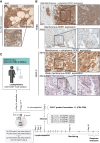
Figure 1
Figure 1
Analysis of ROR1 expression and treatment scheme. ROR1 expression in tumor tissue was assessed by IHC from (A) BM core biopsy of a patient with CLL and (B) needle biopsies from patients with TNBC and NSCLC. Representative patterns of ROR1 expression are shown. Magnification 20×/40×. Scale bars are indicated. C, Patients with ROR1-expressing tumors were enrolled in two treatment arms [CLL (cohort A); TNBC/NSCLC (cohort B)]. Treatment scheme: CD4+ and CD8+ T cells isolated from the leukapheresis products of patients were transduced with an R12(ROR1)/4-1BB/CD3z/EGFRt-expressing CAR vector. IPs were formulated in a 1:1 ratio of CD4+EGFRt+ and CD8+EGFRt+ T cells and administered at indicated dose levels following lymphodepletion with either Cy/Flu or Ox/Cy. Patients were monitored following CAR-T–cell infusion, and the treatment response was evaluated. (C, Created with BioRender.com.)
Figure 2
Figure 2
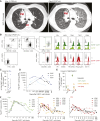
Toxicity and CAR-T–cell kinetics. A, Pre- and posttreatment chest CT images of a patient (X879, cohort B) treated at dose level 4. CT scan was performed 13 days prior to CAR‐T–cell infusion (PRE inf.) and 6 days after CAR‐T–cell infusion (POST inf.). The Pre inf. image shows a large perihilar tumor mass (PM) and lymphangitic spread (LC); the Post inf. image shows a reduction in PM and development of pulmonary infiltrates. B, Phenotype analysis of T cells in the BAL of patient X879 obtained 8 days after CAR-T–cell infusion. CD45+CD3+ T cells were separated into CD4+ and CD8+ T-cell subsets and CAR-T cells identified in these subsets based on the expression of the EGFRt transduction marker. Expressions of PD1, LAG3, TIM3, TIGIT, and CD39 on EGFRt+ and EGFRt cells are shown. Gating of EGFRt positivity was determined based on staining on nontransduced CD45CD3 cells. C, Fraction of EGFRt+ cells per total CD4+ and CD8+ T cells in the peripheral blood of patients in cohort A at the time of CAR-T–cell peak expansion. D, Flap-EF1 copy number per μg DNA isolated from PBMCs of patients in cohort A at the indicated time points after CAR-T–cell infusion. E, Fraction of EGFRt+ cells per total CD4+ and CD8+ T cells in the peripheral blood of patients in cohort B at the time of CAR-T–cell peak expansion. The magnitude of CAR-T–cell expansion was stratified into high (>50% EGFRt+ T cells per total CD8+ T cells), medium (3%–50% EGFRt+ per total CD8+ T cells), and low (<3% EGFRt+ per total CD8+ T cells). F, Flap-EF1 copy number per μg DNA isolated from PBMCs of patients in high-/medium-/low-expanders of cohort B at the indicated time points after CAR-T–cell infusion. Statistical analysis: Data are shown as mean ± SEM (C and E).
Figure 3
Figure 3
Antitumor response and CAR-T–cell infiltration—cohort A. A, Swimmer plot of patients with CLL in cohort A (n = 3). Complete remission (CR), stable disease (SD), partial response (PR), and progressive disease (PD) are indicated. Time point of second CAR‐T–cell infusion (Δ) and occurrence of death (x) are indicated. B, Lymphocyte counts in the peripheral blood of patient with CLL X645 before and after CAR‐T–cell infusion. C, Blood lactate dehydrogenase (LDH) and blood urea nitrogen (BUN) levels of patient with CLL X645 before and after CAR‐T–cell infusion. D, Serum ferritin, C-reactive protein (CRP), and IL-6 in the peripheral blood of patient with CLL X645 in cohort A at the indicated time points before and after CAR‐T–cell infusion (Inf.). E, % EGFRt+ fraction within CD4+ and CD8+ T-cell subsets in the peripheral blood and BM of patient with CLL X714 at indicated time points after first CAR‐T–cell infusion. F, Clinical flow cytometry identifying the frequency of malignant B cells in bone marrow aspirates obtained from patient X714 28 days after the first (F) CAR‐T–cell infusions. CLL cells (bold dots) were identified as CD5+CD19+ cells with low expression of CD20, CD10, CD38, and IgG light chains (κ and λ). G, % EGFRt+ fraction within CD4+ and CD8+ T-cell subsets in the peripheral blood and BM of patient with CLL X714 at indicated time points after second CAR‐T–cell infusion. H, Clinical flow cytometry identifying the frequency of malignant B cells in bone marrow aspirates obtained from patient X714 28 days after the second CAR‐T–cell infusions. CLL cells (bold dots) were identified as CD5+CD19+ cells with low expression of CD20, CD10, CD38, and IgG light chains (κ and λ). I, Dot plots showing the fraction of EGFRt+ CAR-T cells within CD45+CD8+ T cells in the peripheral blood and BM of patient X714 assessed 226 or 393 days after the second infusion. J, Flow cytometry histograms showing the expressions of LAG3, PD1, TIM3, TIGIT, CD39, CD27, CD45RA, and CD45RO within pregated CD8+EGFRt+ CAR-T cells and nontransduced (nTd) CD8+EGFRt bystander T cells in the BM of patient X714 assessed 226 days after the second infusion. K, Section of the archival lung tumor biopsy of patient with CLL X870 in cohort A stained for ROR1 by IHC and counterstained with hematoxylin. Biopsy was obtained 12 months prior to study enrollment. Magnification 20×/40×. Scale bars are indicated. L, % EGFRt+ cells within CD4+ and CD8+ T-cell subsets in the peripheral blood of patient X870.
Figure 4
Figure 4
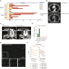
Efficacy and tumor infiltration—cohort B. A, Swimmer plot of patients in cohort B (n = 8 NSCLC; n = 10 TNBC). Duration of stable disease (SD), partial response (PR), and progressive disease (PD) is indicated. Time point of second CAR‐T–cell infusion (Δ) and occurrence of death (x) are specified. B, Pre- and posttreatment chest CT images of patient (X566, cohort B) treated at dose level 1. CT scan was performed 8 days prior to CAR‐T–cell infusion (PRE inf.) and 90 days after CAR‐T–cell infusion (POST inf.). The PRE inf. image shows a large parasternal tumor mass; the Post inf. image shows the reduction of tumor mass. C, Pre- and posttreatment chest CT images of the patient (X461, cohort B) treated at dose level 1. CT scan was performed 8 days prior to CAR‐T–cell infusion (PRE inf.) and 32 days after CAR‐T–cell infusion (POST inf.). The PRE inf. image shows subcutaneous metastatic deposits (top) and neck nodal mass (bottom). D, WPRE copy number per μg DNA extracted from tissue sections of tumor biopsies of cohort B patients (n = 2/3 high-expanders; n = 1/9 medium-expander; n = 4/5 nonexpanders). The time point of tumor biopsy is indicated. E, Immunofluorescence analysis of the tumor biopsy section from patient X552 obtained 38 days after first CAR‐T–cell infusion. Slides were stained for DAPI, CD8, PD1, and TOX. Magnification 20×/40×. Scale bars are indicated. F, ROR1 CAR immunogenicity was assessed by generating effector T-cell lines derived from PBMCs of medium-expanders (n = 4/9) and low-expanders (n = 5/5) obtained 27 days after infusion or later. The killing capacity of effector cell lines was assessed by co-culture with ROR-CAR–positive target cells at indicated (E:T) cell ratios. Mean of technical triplicates and SEM are indicated.

References

    1. Jackson HJ, Rafiq S, Brentjens RJ. Driving CAR T-cells forward. Nat Rev Clin Oncol 2016;13:370–83. - PMC - PubMed
    1. Sadelain M, Rivière I, Riddell S. Therapeutic T cell engineering. Nature 2017;545:423–31. - PMC - PubMed
    1. Raje N, Berdeja J, Lin Y, Siegel D, Jagannath S, Madduri D, et al. . Anti-BCMA CAR T-cell therapy bb2121 in relapsed or refractory multiple myeloma. N Engl J Med 2019;380:1726–37. - PMC - PubMed
    1. Maude SL, Frey N, Shaw PA, Aplenc R, Barrett DM, Bunin NJ, et al. . Chimeric antigen receptor T cells for sustained remissions in leukemia. N Engl J Med 2014;371:1507–17. - PMC - PubMed
    1. Neelapu SS, Locke FL, Bartlett NL, Lekakis LJ, Miklos DB, Jacobson CA, et al. . Axicabtagene ciloleucel CAR T-cell therapy in refractory large B-cell lymphoma. N Engl J Med 2017;377:2531–44. - PMC - PubMed

Publication types

MeSH terms

Substances

LinkOut - more resources