. 2024 Oct 28;19(10):e0309560.
doi: 10.1371/journal.pone.0309560. eCollection 2024.

Novel dual-pathogen multi-epitope mRNA vaccine development for Brucella melitensis and Mycobacterium tuberculosis in silico approach

Affiliations

Novel dual-pathogen multi-epitope mRNA vaccine development for Brucella melitensis and Mycobacterium tuberculosis in silico approach

Yuejie Zhu et al. PLoS One. .

Abstract

Brucellosis and Tuberculosis, both of which are contagious diseases, have presented significant challenges to global public health security in recent years. Delayed treatment can exacerbate the conditions, jeopardizing patient lives. Currently, no vaccine has been approved to prevent these two diseases simultaneously. In contrast to traditional vaccines, mRNA vaccines offer advantages such as high efficacy, rapid development, and low cost, and their applications are gradually expanding. This study aims to develop multi-epitope mRNA vaccines argeting Brucella melitensis and Mycobacterium tuberculosis H37Rv (L4 strain) utilizing immunoinformatics approaches. The proteins Omp25, Omp31, MPT70, and MPT83 from the specified bacteria were selected to identify the predominant T- and B-cell epitopes for immunological analysis. Following a comprehensive evaluation, a vaccine was developed using helper T lymphocyte epitopes, cytotoxic T lymphocyte epitopes, linear B-cell epitopes, and conformational B-cell epitopes. It has been demonstrated that multi-epitope mRNA vaccines exhibit increased antigenicity, non-allergenicity, solubility, and high stability. The findings from molecular docking and molecular dynamics simulation revealed a robust and enduring binding affinity between multi-epitope peptides mRNA vaccines and TLR4. Ultimately, Subsequently, following the optimization of the nucleotide sequence, the codon adaptation index was calculated to be 1.0, along with an average GC content of 54.01%. This indicates that the multi-epitope mRNA vaccines exhibit potential for efficient expression within the Escherichia coli(E. coli) host. Analysis through immune modeling indicates that following administration of the vaccine, there may be variation in immunecell populations associated with both innate and adaptive immune reactions. These types encompass helper T lymphocytes (HTL), cytotoxic T lymphocytes (CTL), regulatory T lymphocytes, natural killer cells, dendritic cells and various immune cell subsets. In summary, the results suggest that the newly created multi-epitope mRNA vaccine exhibits favorable attributes, offering novel insights and a conceptual foundation for potential progress in vaccine development.


Conflict of interest statement

NO authors have competing interests.

Figures

Fig 1
Fig 1
(A) Prediction of subcellular localization of protein Omp25. The pink part indicates the extracellular domain. (B) Prediction of subcellular localization of protein Omp31. (C) Prediction of subcellular localization of protein MPT70. (D) Prediction of subcellular localization of protein MPT83. (E) The N-terminal region of the signal peptide. Reported for Sec/SPI, Sec/SPII, Tat/SPI, and Tat/SPII. The signal peptide sequence of Omp25: MRTLKSLVIVSAALLPFSATAFAA. (F) The signal peptide sequence of Omp31: MKSVILASIAAMFATSAMAA. (G) Signal peptide sequence of MPT70: MKVKNTIAATSFAAAGLAALAVAVSPPAAA. (H) Signal peptide sequence of MPT83: MINVQAKPAAAASLAAIAIAFLAGC.
Fig 2
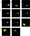
Fig 2. Conformational B cell epitope 3D map.
(A-C)OMP25 protein B cell conformational epitope.(D-H)OMP30 protein B cell conformational epitope.(I)MPT70protein B cell conformational epitope.(G-K)MPT83protein B cell conformational epitope.
Fig 3
Fig 3. The final HTL epitopes, CTL epitopes, LB epitopes, and CB epitopes were selected for MEV design.
As illustrated in the figure, “green” stands for the T-cell epitope of Omp25, “blue” stands for the T-cell epitope of Omp31, “purple” stands for the T-cell epitope of MPT70, and “red” stands for the T-cell epitope of MPT83.
Fig 4
Fig 4
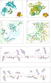
(A) Representative lateral views of HLA-A*02:01 interacting with CTL epitopes. (B) Representative lateral views of HLA-DRB1*01:01 interacting with HTL epitopes. (C) The interaction interface residues between HLA-A*02:01 and CTL epitopes were analyzed by PyMol. (D) The interaction interface residues between HLA-DRB1*01:01 and HTL epitopes were analyzed by PyMol. (E) The interaction interface residues between HLA-A*02:01 and CTL epitopes were analyzed by Ligplot. (F) The interaction interface residues between HLA-DRB1*01:01 and HTL epitopes were analyzed by Ligplot.
Fig 5
Fig 5
(A) The 3D structure of Omp25. (B) The 3D structure of Omp31. (C) The 3D structure of MPT70. (D) The 3D structure of MPT83. (E) The final structure diagram of the mRNA vaccines. Multi-epitope peptides(deep yellow), EAAAK(Pale pink), HBDC(faint yellow)are adjuvants.
Fig 6
Fig 6
(A) The secondary structure of the mRNA vaccine was predicted by NetSurfP-2.0. With a threshold of 25%, red upward elevation implies residue exposure, whereas sky blue denotes buried residue. As illustrated in the figure, “orange” stands for the alpha-helix, “blue” stands for the β-fold, and “purple” stands for the random curl. Thereinto, the disorder is represented as a bloated black line, with the thickness of the line equaling the probability of disordered residue. (B) The secondary structure of the mRNA vaccine was predicted by SOPMA. (C) The tertiary structure of the mRNA vaccine was predicted by AlphaFold.
Fig 7
Fig 7
(A) Z score of the model of the mRNA vaccine model developed by ProSA-web. (B) Ramachandran plots of the mRNA vaccine. As illustrated in the figure, “dark green” stands for the most favored regions, “green” stands for the additional allowed regions, “light green” stands for the generously allowed regions, and “white” stands for the disallowed regions.
Fig 8
Fig 8
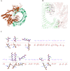
(A) The docking model of mRNA vaccine -TLR4 predicted by HDOCK. (B) The interaction interface residues between mRNA vaccine and TLR4 were analyzed by PyMol. (C) The interaction interface residues between mRNA vaccine and TLR4 were analyzed by Ligplot.
Fig 9
Fig 9
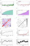
(A) The deformability plot represents the complexes formed by the combination of mRNA vaccine with TLR4. (B) The B-factor value plot represents the complexes formed by the combination of mRNA vaccine with TLR4. (C) The variance plot represents the complexes formed by the combination of mRNA vaccine with TLR4. (D) The eigenvalue plot represents the complexes formed by the combination of mRNA vaccine with TLR4. (E) The covariance matrix represents the complexes formed by the combination of mRNA vaccine with TLR4. As illustrated in the figure, “red” stands for the correlated motions, “white” stands for the uncorrelated motions, and “blue” stands for the anti-correlated motions. (F) The cross-correlation matrix diagram represents the complexes formed by the combination of mRNA vaccine with TLR4. (G) Root-mean-square deviation (RMSD) of the mRNA vaccine -TLR4. RMSD over the entire simulation, where the ordinate is the value of RMSD, and the abscissa is time (ns). (H) RMSF values of the mRNA vaccine -TLR4 over the entire simulation, where the ordinate is RMSF, and the abscissa is residue. (I) The radius of gyration (Rg) over the entire simulation, where the ordinate is Rg, and the abscissa is time (ns). (F) The number of total hydrogen bonds versus simulation time.
Fig 10
Fig 10. Projection of trajectories into PC1 and PC2 for the mRNA vaccine -TLR4 complex.
Fig 11
Fig 11
(A) The level of immunoglobulins after vaccination. As illustrated in the figure, “yellow” stands for the IgM+IgG, “green” stands for the IgM, “blue” stands for the IgG1+IgG2, “purple” stands for the IgG1, and “red” stands for the IgG2. (B) The concentration of cytokines and interleukins after vaccination. As illustrated in the figure, “D” stands for the danger signal. (C) The level of B-cell populations after vaccination. As illustrated in the figure, “black” stands for the total number of B cells, and “green” stands for the memory B cell. (D) B lymphocytes population per entity-state (i.e., showing counts for active, presenting on class-II, internalized the ag, duplicating, and anergic. (E) The level of plasma B lymphocytes after vaccination. (F) The level of CD4 T-helper lymphocytes after vaccination. (G) The level of CD4 T-regulatory lymphocytes after vaccination. Both total, memory, and per entity-state counts are plotted here. (H) The level of CD8 T-cytotoxic lymphocytes after vaccination. (I) The level of natural killer cells after vaccination. (J) The level of dendritic cells after vaccination. The curves show the total number broken down to active, resting, internalized, and presenting the ag. (K) The level of macrophages after vaccination. (L) The level of epithelial cells after vaccination.
Fig 12
Fig 12. RNA secondary structure.
(A) The optimal secondary structure of mRNA vaccine has the minimum free energy. (B) The centroid structure of mRNA vaccine.
Fig 13
Fig 13
(A) The calculation of the codon adaptation index. (B) In silico cloning of the mRNA vaccine optimized codons (purple) into pET-28a (+) expression vector between XhoI and BamHI restriction sites.

Similar articles

Cited by

References

    1. Zhou X, Li X, Wu M. miRNAs reshape immunity and inflammatory responses in bacterial infection. Signal Transduct Target Ther. 2018;3:14. doi: 10.1038/s41392-018-0006-9 - DOI - PMC - PubMed
    1. Zhuge X, Sun Y, Jiang M, Wang J, Tang F, Xue F, et al.. Acetate metabolic requirement of avian pathogenic Escherichia coli promotes its intracellular proliferation within macrophage. Vet Res. 2019;50(1):31. doi: 10.1186/s13567-019-0650-2 - DOI - PMC - PubMed
    1. Paul S, Peddayelachagiri BV, Gogoi M, Nagaraj S, Ramlal S, Konduru B, Batra HV. Genome-wide unique insertion sequences among five Brucella species and demonstration of differential identification of Brucella by multiplex PCR assay. Sci Rep. 2020;10(1):6368. doi: 10.1038/s41598-020-62472-3 - DOI - PMC - PubMed
    1. Rutaihwa LK, Sasamalo M, Jaleco A, Hella J, Kingazi A, Kamwela L, et al.. Insights into the genetic diversity of Mycobacterium tuberculosis in Tanzania. PLoS One. 2019;14(4):e0206334. doi: 10.1371/journal.pone.0206334 - DOI - PMC - PubMed
    1. Nandini P, Jakka P, Murugan S, Mazumdar V, Kumar D, Prakash R, et al.. Immuno-profiling of Brucella proteins for developing improved vaccines and DIVA capable serodiagnostic assays for brucellosis. Front Microbiol. 2023;14:1253349. doi: 10.3389/fmicb.2023.1253349 - DOI - PMC - PubMed

MeSH terms

LinkOut - more resources