. 2024 Oct 28;20(10):e1012650.
doi: 10.1371/journal.ppat.1012650. eCollection 2024 Oct.

Delineating the functional activity of antibodies with cross-reactivity to SARS-CoV-2, SARS-CoV-1 and related sarbecoviruses

Affiliations

Delineating the functional activity of antibodies with cross-reactivity to SARS-CoV-2, SARS-CoV-1 and related sarbecoviruses

Felicitas Ruiz et al. PLoS Pathog. .

Abstract

The recurring spillover of pathogenic coronaviruses and demonstrated capacity of sarbecoviruses, such SARS-CoV-2, to rapidly evolve in humans underscores the need to better understand immune responses to this virus family. For this purpose, we characterized the functional breadth and potency of antibodies targeting the receptor binding domain (RBD) of the spike glycoprotein that exhibited cross-reactivity against SARS-CoV-2 variants, SARS-CoV-1 and sarbecoviruses from diverse clades and animal origins with spillover potential. One neutralizing antibody, C68.61, showed remarkable neutralization breadth against both SARS-CoV-2 variants and viruses from different sarbecovirus clades. C68.61, which targets a conserved RBD class 5 epitope, did not select for escape variants of SARS-CoV-2 or SARS-CoV-1 in culture nor have predicted escape variants among circulating SARS-CoV-2 strains, suggesting this epitope is functionally constrained. We identified 11 additional SARS-CoV-2/SARS-CoV-1 cross-reactive antibodies that target the more sequence conserved class 4 and class 5 epitopes within RBD that show activity against a subset of diverse sarbecoviruses with one antibody binding every single sarbecovirus RBD tested. A subset of these antibodies exhibited Fc-mediated effector functions as potent as antibodies that impact infection outcome in animal models. Thus, our study identified antibodies targeting conserved regions across SARS-CoV-2 variants and sarbecoviruses that may serve as therapeutics for pandemic preparedness as well as blueprints for the design of immunogens capable of eliciting cross-neutralizing responses.


Conflict of interest statement

J.O. is a consultant for Aerium Therapeutics, Inc. J.O. and J.G are listed on a patent application (22-173-US-PSP2) and license agreement with Aerium Therapeutics, Inc. for C68.61. H.Y.C reported consulting with Ellume, Merck, Abbvie, Pfizer, Medscape, Vindico, and the Bill and Melinda Gates Foundation. She has received research funding from Gates Ventures, and support and reagents from Ellume and Cepheid outside of the submitted work. T.N.S. consults for Apriori Bio and Vir Biotechnology on deep mutational scanning. The lab of T.N.S. has received sponsored research agreements unrelated to the present work from Vir Biotechnology, Aerium Therapeutics, Inc. and Invivyd, Inc.

Figures
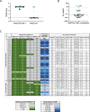
Fig 1
Fig 1. Identification and molecular characteristics of SARS-CoV-1 and SARS-CoV-2 cross-reactive mAbs.
(A) C68 mAbs binding to SARS-CoV-2 or SARS-CoV-1 recombinant spike glycoprotein by ELISA (OD450nm values). C68 antibodies that bound SARS-CoV-2 trimer and SARS-CoV-1 trimer (designated by green closed circles; SARS-CoV-2 trimer-specific mAbs shown in black closed circles). Antibodies tested at 500 ng/ml, represents the average of two technical replicates shown as background-corrected OD450nm values. Dotted line represents 3 standard deviations above the negative control Fl6v3, [43] an influenza-specific mAb. (B) Neutralization of spike-pseudotyped lentiviruses by C68 mAbs against SARS-CoV-2 WH-1. C68 antibodies that bound SARS-CoV-2 trimer and SARS-CoV-1 trimer are indicated (in green). IC50 values (μg/mL) were calculated by nonlinear regression analysis in the statistical software package PRISM from at least two independent experiments. Black dotted line represents the median IC50 value for the SARS-CoV-2 specific mAbs. Green line represents the median IC50 value for the cross-reactive mAbs. (C) Summary table representing gene family usage, somatic hypermutation percent (% SHM relative to germline), and a heatmap of functional activity of C68 mAbs. OD450 values indicate absorbance at OD450nm values where each mAb was tested at 500 ng/ml from technical replicates and background-corrected.
Fig 2
Fig 2. Comprehensive functional characterization of C68 cross-reactive mAbs.
(A) C68 mAbs binding to SARS-CoV-2 variants and SARS-CoV-1 recombinant spike glycoprotein by ELISA (OD450nm values). Half-maximal effective concentrations (EC50 values) calculated by nonlinear regression analysis from at least two independent replicates. *Binding activity for C68.61 from [31]. (B) SARS-CoV-2 variants and sarbecovirus neutralization activity. IC50 values (μg/mL) were calculated by nonlinear regression analysis from at least two independent experiments with technical replicates. Samples with IC50 values above 20 μg/mL were plotted at 20 μg/mL (no activity). *Neutralization activity for C68.61 and S309 against SARS-CoV-2 variants and SARS-CoV-1 from [31]. S309 neutralizing activity against WH-1 was not determined (ND). (C) Pan-sarbecovirus RBD binding of select mAbs. Each antibody (row) was experimentally measured for binding to each sarbecovirus RBD (column) via a multiplexed FACS-seq titration assay with a yeast-display library of sarbecovirus RBDs. EC50 (ng/ml) value represents the geometric mean across internally replicated barcodes within the library linked to the same sarbecovirus RBD. (D) C68 mAbs can mediate antibody dependent cellular cytotoxicity. Cells expressing SARS-CoV-2 D614G spike cells were incubated with C68 mAbs and effector cells (PBMC) for 4h. NK cell degranulation (% CD107a+) was measured as a proxy for ADCC. The assay was run in technical duplicate with two independent PBMC donors. Background subtracted values (target cells + PBMC only) from a single donor are shown.
Fig 3
Fig 3. Epitope profiling by deep mutational scanning identifies a group of C68 mAbs targeting class 4 epitopes.
(A) Complete escape maps of antibody-binding escape mutations for C68 mAbs using a yeast-displayed SARS-CoV-2 RBD deep mutational scanning system (Wuhan-Hu-1 RBD DMS for C68.88 and C68.239; Omicron BA.2 RBD DMS for C68. 83, C68.121, C68.348). Residue colors are assigned based on effect these mutations have on RBD expression (with yellow indicating mutations deleterious for RBD expression). The height of letters in the logo plots indicate level of escape by that amino acid at that site. Logo plot residue numbering is based on SARS-CoV-2 WH-1 numbering. Experiments were performed in biological duplicate using independent mutant RBD libraries [14] that correlate well (S10B Fig) so escape fractions represent the average of these two independent biological replicates. On the right, sites of escape for C68 mAbs are mapped onto the RBD structure and gradient of red coloring indicates magnitude of escape fraction at each indicated site. (B) Multiple sequence alignments of select SARS-CoV-2 variants and sarbecoviruses are shown corresponding to sites of escape. Residue numbering based on SARS-CoV-2 Wuhan-Hu-1 sequence with shared amino acids denoted by dots (.) and differences across sarbecoviruses relative to the reference sequence (SARS-CoV-2 Wuhan-Hu-1) indicated. Pink triangles depict the sites of escape identified by the escape maps (Fig 3A).
Fig 4
Fig 4. C68 mAbs target class 5 epitopes that are relatively invariant regions on the Spike Protein.
(A) Complete escape maps of antibody-binding escape mutations for C68 mAbs using a yeast-displayed SARS-CoV-2 RBD deep mutational scanning system (Omicron BA.2 RBD DMS for all class 5 mAbs). Residue colors are assigned based on effect these mutations have on RBD expression. The height of letters in the logo plots indicate escape scores. Logo plot residue numbering is based on SARS-CoV-2 Omicron BA.2. Experiments were performed in biological duplicate using independent mutant RBD libraries [14] that correlate well (S10B Fig) so escape fractions represent the average of these two independent biological replicates. On the right, sites of escape for C68 mAbs are mapped onto the RBD structure and gradient of red coloring indicates magnitude of escape fraction at each indicated site. (B) Multiple sequence alignments of select SARS-CoV-2 variants and sarbecoviruses are shown corresponding to sites of escape. Residue numbering based on SARS-CoV-2 Wuhan-Hu-1 sequence with shared amino acids denoted by dots (.) and differences across sarbecoviruses relative to the reference sequence (SARS-CoV-2 Wuhan-Hu-1) indicated. Pink triangles depict the sites of escape identified by the escape maps (Fig 3A).
Fig 5
Fig 5. Selection experiments to identify escape mutation in the presence of SARS-Cov-1 and SARS-CoV-2 encoding VSV in cell culture and SARS-CoV-2 evolution observed in nature.
(A) C68 mAbs selection experiments in the context of SARS-CoV-1 replication competent VSV-G. Frequency of variants in viral population in the presence of C68 mAbs (variant defined as occurring at a frequency greater than 3%). (B) Neutralization activity of C68 mAbs (C68.61 in red and C68.185 in blue) and S309 (in grey) against recently circulating and dominant SARS-CoV-2 variant spike-pseudotyped lentiviruses. Antibodies were two-fold serially diluted and tested starting at 20 and/or 30 μg/mL. Samples with IC50 values above the tested concentration were plotted at that concentration (>20 μg/mL is no activity). (C) Phylogeny of SARS-CoV-2 Pangolin clade sequences containing C68 mAb RBD binding escape mutations (from Fig 4A) (Nextstrain) for C68.61 (top panel) and C68.185 (bottom panel). Circles in teal are the residues found in SARS-CoV-2 Wuhan-Hu-1 whereas viral genomes containing escape mutations are indicated by yellow/blue circles. Depicts 3900 SARS-CoV-2 viral genomes that are sampled between December 2019 and March 2024. (Nextstrain: https://nextstrain.org, CC-BY-4.0 license).

Update of

References

    1. Ksiazek TG, Erdman D, Goldsmith CS, Zaki SR, Peret T, Emery S, et al.. A novel coronavirus associated with severe acute respiratory syndrome. N Engl J Med. 2003. May 15;348(20):1953–66. doi: 10.1056/NEJMoa030781 - DOI - PubMed
    1. Drosten C, Günther S, Preiser W, van der Werf S, Brodt H-R, Becker S, et al.. Identification of a novel coronavirus in patients with severe acute respiratory syndrome. N Engl J Med. 2003. May 15;348(20):1967–76. doi: 10.1056/NEJMoa030747 - DOI - PubMed
    1. Zaki AM, van Boheemen S, Bestebroer TM, Osterhaus ADME, Fouchier RAM. Isolation of a novel coronavirus from a man with pneumonia in Saudi Arabia. N Engl J Med. 2012. Nov 8;367(19):1814–20. - PubMed
    1. Zhou P, Yang X-L, Wang X-G, Hu B, Zhang L, Zhang W, et al.. A pneumonia outbreak associated with a new coronavirus of probable bat origin. Nature. 2020. Mar;579(7798):270–3. doi: 10.1038/s41586-020-2012-7 - DOI - PMC - PubMed
    1. Walls AC, Park Y-J, Tortorici MA, Wall A, McGuire AT, Veesler D. Structure, Function, and Antigenicity of the SARS-CoV-2 Spike Glycoprotein. Cell. 2020. Apr 16;181(2):281–292.e6. doi: 10.1016/j.cell.2020.02.058 - DOI - PMC - PubMed

MeSH terms

Supplementary concepts