. 2025 Jan;12(1):e2405597.
doi: 10.1002/advs.202405597. Epub 2024 Oct 28.

A Cardiac-Targeting and Anchoring Bimetallic Cluster Nanozyme Alleviates Chemotherapy-Induced Cardiac Ferroptosis and PANoptosis

Affiliations

A Cardiac-Targeting and Anchoring Bimetallic Cluster Nanozyme Alleviates Chemotherapy-Induced Cardiac Ferroptosis and PANoptosis

Junyue Xing et al. Adv Sci (Weinh). 2025 Jan.

Abstract

Doxorubicin (DOX), a potent antineoplastic agent, is commonly associated with cardiotoxicity, necessitating the development of strategies to reduce its adverse effects on cardiac function. Previous research has demonstrated a strong correlation between DOX-induced cardiotoxicity and the activation of oxidative stress pathways. This work introduces a novel antioxidant therapeutic approach, utilizing libraries of tannic acid and N-acetyl-L-cysteine-protected bimetallic cluster nanozymes. Through extensive screening for antioxidative enzyme-like activity, an optimal bimetallic nanozyme (AuRu) is identified that possess remarkable antioxidant characteristics, mimicking catalase-like enzymes. Theoretical calculations reveal the surface interactions of the prepared nanozymes that simulate the hydrogen peroxide decomposition process, showing that these bimetallic nanozymes readily undergo OH⁻ adsorption and O₂ desorption. To enhance cardiac targeting, the atrial natriuretic peptide is conjugated to the AuRu nanozyme. These cardiac-targeted bimetallic cluster nanozymes, with their anchoring capability, effectively reduce DOX-induced cardiomyocyte ferroptosis and PANoptosis without compromising tumor treatment efficacy. Thus, the therapeutic approach demonstrates significant reductions in chemotherapy-induced cardiac cell death and improvements in cardiac function, accompanied by exceptional in vivo biocompatibility and stability. This study presents a promising avenue for preventing chemotherapy-induced cardiotoxicity, offering potential clinical benefits for cancer patients.

Keywords: DOX‐induced cardiotoxicity; PANoptosis; antioxidant activity; bimetallic cluster nanozyme; ferroptosis.


Conflict of interest statement

The authors declare no conflict of interest.

Figures

Scheme 1
Scheme 1
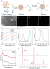
Schematic illustration of a) preparation of bimetallic cluster nanozyme library and cardiac‐targeted ATBMzyme and b) application for the treatment of DIC by the cardiac‐targeted ATBMzyme.
Figure 1
Figure 1
Preparation and screen of bimetallic nanozymes. A) Schematic representation of the preparation of different bimetallic nanozymes. B) The CAT‐like activity of different bimetallic nanozymes. C) The SOD‐like activity of different bimetallic nanozymes. D) ABTS radical scavenging ratio of different bimetallic nanozymes. E) DPPH radical scavenging ratio of different bimetallic nanozymes. F) Radar chart (SOD/CAT‐like activity, ABTS, and DPPH radical scavenging of different bimetallic nanozymes. G) Surface configurations of different modeled initial, transition, and final states for the simulated CAT‐like catalytic processes.
Figure 2
Figure 2
Preparation and characterization of nanozymes. A) Scheme representing the preparation of ATBMzyme. B) TEM image of ATBMzyme (Scale bar = 20 nm, inset scale bar of TEM is 2 nm). C–E) Elemental mapping of ATBMzyme. F) FTIR of BMzyme, TBMzyme, and ATBMzyme. G) XPS spectra of Au element in ATBMzyme. H) XPS spectra of Ru 3d in ATBMzyme. I) PXRD of BMzyme and TBMzyme. J) 1H NMR of TBMzyme, and ATBMzyme.
Figure 3
Figure 3
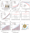
Analysis of the ROS scavenging potential of nanozyme. A) UV–vis spectrum of BMzyme, TBMzyme, and ATBMzyme in buffer solution. B) SOD‐like activities of BMzyme, TBMzyme, and ATBMzyme after incubation for different concentration in buffer solution (pH 7.4). C) The decomposition of H2O2 by BMzyme and TBMzyme was assessed after incubation in a buffer solution at pH 7.4 by monitoring absorbance changes at 240 nm. D) CAT‐like activities of BMzyme, TBMzyme, and ATBMzyme after incubation in buffer solution (pH 7.4) for various durations. E,F) ATBMzyme reduces the generation of superoxide radical (·O2 ) and hydroxyl radical (·OH), illustrated by ESR spectroscopy. G) ABTS•+ scavenging rate of BMzyme, TBMzyme, ATBMzyme. H) DPPH• scavenging rate of BMzyme, TBMzyme, ATBMzyme. I) Schematic representation of cascade ROS and RNS scavenging activities of ATBMzyme. n = 3, data represent means ± SD.
Figure 4
Figure 4
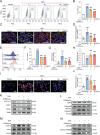
ATBMzyme significantly alleviates Doxorubicin‐induced ferroptosis and PANoptosis in vitro. AC16 cells were pre‐treated with ATBMzyme for 20 h, then exposed to DOX (800 nm) for 24 h. The cells were subsequently collected for the following experiments A) Flow cytometric calculation of the percentage of PI+ AC16 cells (n = 6). B) Quantification of PI staining of AC16 (n = 6). C) Representative micrographs of mitochondrial membrane potential (JC‐1 staining) in AC16, Green: JC‐1 monomer; Red: JC‐1 aggregates (scale bars, 50 µm; n = 6). D) Quantification of JC‐1 staining in AC16 (n = 6). E) Representative flow cytometry graphs of intracellular ROS (DCFH‐DA staining) in AC16 (n = 6). F) Quantification of intracellular ROS (DCFH‐DA staining) in AC16 (n = 6). G) Relative MDA levels in AC16 (n = 6). H) GSH/GSSH ratio in AC16 (n = 6). I) Representative micrographs of intracellular iron level (FerroOrange staining) in AC16 (scale bars, 50 µm; n = 6). J) Quantification of FerroOrange staining of AC16 (n = 6). K) Representative immunoblots of ferroptosis‐related proteins in AC16 (n = 6). L–N) Representative immunoblots of PANoptosis‐related proteins in AC16 (n = 6). Data has been expressed as mean ± SD and analyzed using one‐way ANOVA followed by Tukey's post hoc test, *** p < 0.001 versus the Vehicle group; ### p < 0.001 versus the DOX group.
Figure 5
Figure 5
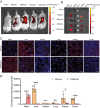
Heart‐specific targeting ability of BMzyme and ATBMzyme. A) Representative image of live imaging of animals. B) Representative fluorescent image of heart, liver, spleen, lung, kidney and tumor. C) Representative immunofluorescence images, and D) fluorescence intensity quantification of the distribution of BMzyme and ATBMzyme in the tissues from different organs (scale bars, 50 µm; n = 6). Data were expressed as mean ± SD and analyzed using a two‐tailed Student's t‐test, * p < 0.05, ** p < 0.01, *** p < 0.001.
Figure 6
Figure 6
Cardiac‐targeting ATBMzyme significantly alleviates Doxorubicin‐induced myocardial injury and reduces cardiac function. A) Experiment design: A chronic DIC mouse model was established in Babl/c mice bearing 4T1 tumor cells, BMzyme and ATBMzyme were used for the treatment of the DIC model. All tests and analyses were implemented 1 week after the last DOX injection. B) Heart weight/Tibial length ratio (n = 6). C) Representative echocardiographic images of cardiac function (scale bar = 300 ms, n = 6). D,E) Quantitative analysis of ejection fraction and fraction shortening. (n = 6). F–I) Serum levels of lactate dehydrogenase (LDH), cardiac troponin T (cTnT), creatine kinase‐myocardial band (CK‐MB), and N‐terminal pro‐B type natriuretic peptide (NT‐proBNP) to assess cardiac injury (n = 6). J) HE and Masson staining of heart from Figure 6A, scale bar were indicated in the Figure. Data were expressed as mean ± SD and analyzed using one‐way ANOVA followed by Tukey's post hoc test, ** p < 0.01 and *** p < 0.001 versus Vehicle group; #p < 0.05, ## p < 0.01 and ### p < 0.001 versus DOX group.
Figure 7
Figure 7
The cardiac‐targeting ATBMzyme significantly alleviates DIC by preventing ferroptosis‐induced myocardial cell death. A) Representative micrographs of heart tissues examined by transmission electron microscopy (scale bars, 2 µm). B) Representative micrographs of cardiac intracellular ROS levels (DHE staining) (scale bars, 50 µm; n = 6). C) Quantification of DHE staining in sections of heart tissues (n = 6). D) Representative immunohistochemistry micrographs of cardiac 4‐HNE staining (scale bars, 50 µm; n = 6). E) Quantification of 4‐HNE staining in heart sections (n = 6). F) Representative immunoblots of ferroptosis–related proteins in heart tissues (n = 6). G) Quantitative analysis of ferroptosis‐related proteins in heart tissues (n = 6). Data were expressed as mean ± SD and analyzed using one‐way ANOVA followed by Tukey's post hoc test, *** p < 0.001 versus Vehicle group; # p < 0.05, ## p < 0.01, ### p < 0.001 versus DOX group.
Figure 8
Figure 8
Cardiac‐targeting ATBMzyme significantly alleviates DIC, by preventing PANoptosis‐induced myocardial cell death. A) Representative images of TUNEL staining in heart sections, green fluorescence represents the TUNEL‐positive cells (scale bars, 50 µm; n = 6). B) Quantification of TUNEL staining in heart sections (n = 6). C–E) Representative immunoblots of PANoptosis‐related proteins in heart tissues (n = 6). F) Quantitative analysis of PANoptosis‐related proteins in heart tissues (n = 6). G) Schematic diagram depicting how ATBMzyme alleviates chemotherapy‐induced cardiac ferroptosis and PANoptosis. The data were expressed as mean ± SD and analyzed using one‐way ANOVA followed by Tukey's post hoc test, * p<0.05, ** p < 0.01, *** p < 0.001 versus Vehicle group; ## p < 0.01, ### p < 0.001 versus DOX group.
Figure 9
Figure 9
Both BMzyme and ATBMzyme show protection against DOX‐induced renal and liver damage. A) Representative micrographs of H&E staining in liver and kidney sections (scale bars, 50 µm; n = 6). B) Representative images of TUNEL staining in hepatic and renal sections, green fluorescence represents the TUNEL‐positive cells (scale bars, 50 µm; n = 6). Quantification of TUNEL staining in kidney C) and liver D). E–H) Serum levels of alanine aminotransferase (ALT), aspartate aminotransferase (AST), urea nitrogen (UREA), and creatinine (CREA) to assess hepatic and renal injury (n = 6). The data were expressed as mean ± SD and analyzed using one‐way ANOVA followed by Tukey's post hoc test, ** p < 0.01 and *** p < 0.001 versus Vehicle group; # p < 0.05, ## p < 0.01, and ### p < 0.001 versus DOX group.

References

    1. Jamaledin R., Yiu C. K. Y., Zare E. N., Niu L. N., Vecchione R., Chen G., Gu Z., Tay F. R., Makvandi P., Adv. Mater. 2020, 32, 2002129. - PubMed
    1. Lena A., Wilkenshoff U., Hadzibegovic S., Porthun J., Rösnick L., Fröhlich A. K., Zeller T., Karakas M., Keller U., Ahn J., Bullinger L., Riess H., Rosen S. D., Lyon A. R., Lüscher T. F., Totzeck M., Rassaf T., Burkhoff D., Mehra M. R., Bax J. J., Butler J., Edelmann F., Haverkamp W., Anker S. D., Packer M., Coats A. J. S., von Haehling S., Landmesser U., Anker M. S., J. Am Coll. Cardiol. 2023, 81, 1569. - PubMed
    1. Ewer M. S., Ewer S. M., Nat. Rev. Cardiol. 2015, 12, 547. - PubMed
    1. Fan M., Li H., Shen D., Wang Z., Liu H., Zhu D., Wang Z., Li L., Popowski K. D., Ou C., Zhang K., Zhang J., Cheng K., Li Z., Adv. Sci. 2022, 9, e2203505. - PMC - PubMed
    1. Sawicki K. T., Sala V., Prever L., Hirsch E., Ardehali H., Ghigo A., Annu. Rev. Pharmacol. Toxicol. 2021, 61, 309. - PubMed

LinkOut - more resources