. 2024 Dec;120(5):2151-2171.
doi: 10.1111/tpj.17109. Epub 2024 Oct 28.

Targeted modulation of pennycress lipid droplet proteins impacts droplet morphology and seed oil content

Affiliations

Targeted modulation of pennycress lipid droplet proteins impacts droplet morphology and seed oil content

Athanas Guzha et al. Plant J. 2024 Dec.

Abstract

Lipid droplets (LDs) are unusual organelles that have a phospholipid monolayer surface and a hydrophobic matrix. In oilseeds, this matrix is nearly always composed of triacylglycerols (TGs) for efficient storage of carbon and energy. Various proteins play a role in their assembly, stability and turnover, and even though the major structural oleosin proteins in seed LDs have been known for decades, the factors influencing LD formation and dynamics are still being uncovered mostly in the "model oilseed" Arabidopsis. Here we identified several key LD biogenesis proteins in the seeds of pennycress, a potential biofuel crop, that were correlated previously with seed oil content and characterized here for their participation in LD formation in transient expression assays and stable transgenics. One pennycress protein, the lipid droplet associated protein-interacting protein (LDIP), was able to functionally complement the Arabidopsis ldip mutant, emphasizing the close conservation of lipid storage among these two Brassicas. Moreover, loss-of-function ldip mutants in pennycress exhibited increased seed oil content without compromising plant growth, raising the possibility that LDIP or other LD biogenesis factors may be suitable targets for improving yields in oilseed crops more broadly.

Keywords: arabidopsis thaliana; endoplasmic reticulum; lipid droplet proteins; lipid droplets; pennycress; seeds; seipin; thlaspi arvense; triacylglycerols.


Conflict of interest statement

None declared.

Figures

Figure 1
Figure 1
Different lipid related genes are more expressed with increased maturity of pennycress embryos. (A) Heat map diagram showing relative transcript abundance of some pennycress lipid associated genes in the developing embryos of two pennycress accessions. The expression levels of selected candidates were measured using three replicates of developing pennycress embryos at 10, 14, 17, and 20 days post anthesis (DPA) using a high oil accession (TAMN106) and a low oil accession (Ames 32872). Red, positive log fold‐change (log FC) indicates higher expression in the embryo; green, negative log FC. (B) Representative confocal laser‐scanning microscopy images of BODIPY‐stained LDs (green) in cotyledon tissues of pennycress MN106 embryos at 10, 14, 17, and 20 DPA. Blue is autofluorescence from chlorophyll. Bar = 5 μm.
Figure 2
Figure 2
Coding DNA sequences of the four pennycress LD associated proteins (SEIPIN1, LDAP1, LDAP2, and LDAP3) with an eGFP tag were cloned and expressed in Nicotiana benthamiana leaves for localization. (A–D) Representative confocal laser scanning microscopy (Z‐sections) of N. benthamiana leaves transformed with SEIPIN1 (A), LDAP1 (B), LDAP2 (C), and LDAP3 (D) shows that the proteins localize to LDs (arrows). The LDs were stained with Nile red, while HDEL‐CFP (cyan) was used as an ER marker. Blue is autofluorescence from the chloroplasts. Images are representative for at least ten images from nine leaves derived from three independent transformations for each protein. Bars = 10 μm.
Figure 3
Figure 3
Lipid droplet abundance in Nicotiana benthamiana leaves infiltrated with LD associated proteins. (A–D) Representative confocal laser scanning microscopy images (z‐stacks) of LD abundance in N. benthamiana leaves showing that the ectopic overexpression of four pennycress LD associated proteins: Seipin1 (A), LDAP1 (B), LDAP2 (C), and LDAP3 (D) coding sequences resulted in the alteration of LD abundance. LDs are stained (green) with BODIPY while blue is autofluorescence from the chloroplasts. Bars = 20 μm. (E–H) The LD abundance in infiltrated N. benthamiana leaves was quantified. Values are averages of three individual experiments with at least three images for each replicate. Error bars show SD. Statistical differences to the mock were determined for each construct (*P < 0.05; **P < 0.01; ***P < 0.001; ****P < 0.0001, Kruskal–Wallis test followed by Dunn's test). (I–L) Neutral lipid content in the N. benthamiana leaves transformed with the four proteins is also quantified. Values are averages from three leaves per line. Error bars show SD. Statistical differences to the mock were determined for each construct (*P < 0.05; **P < 0.01, ***P < 0.001, ****P < 0.0001, Kruskal–Wallis test followed by Dunn's test).
Figure 4
Figure 4
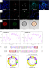
Pennycress LDIP localizes to LDs. (A–C) (A) Representative confocal laser scanning microscopy images showing that the coding DNA sequence of pennycress LDIP with an eGFP tag localizes to the LDs stained with Nile red (arrows) when transiently expressed in Nicotiana benthamiana leaves. Blue is chlorophyll autofluorescence. CFP‐HDEL (cyan) was used as the ER marker. The images are individual optical sections. Bar = 20 μm. Similarly, heterologous expression of eGFP tagged pennycress LDIP in Arabidopsis leaves (B), bar = 20 μm, and pollen grains (C) shows a LD localization. Bar = 5 μm. (D, E) Hydropathy plots derived from amino acid sequence of pennycress LDIP (D) and Arabidopsis LDIP (E) based on the Kyte‐Doolittle scale. Note the presence of a hydrophobic region in the middle of the polypeptide sequence of both proteins. A hydrophobic region is also present toward the N‐terminus around the 50th amino acid residue on both sequences. (F) The N‐terminus hydrophobic region is conserved in the two proteins and the red box highlights the region predicted to form an amphipathic α‐helix. (G) Helical wheel projection of amino acid residues 55–72 and 48–65 in pennycress LDIP and Arabidopsis LDIP, respectively show that these regions form an amphipathic α‐helix. Hydrophobic amino acid residues are colored yellow. The direction of the arrowhead in the helical wheel indicates the position of the hydrophobic face along the axis of the helix and its predicted to form an α‐helix.
Figure 5
Figure 5
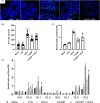
Ectopic expression of pennycress LDIP induces an accumulation of storage lipids in Nicotiana benthamiana leaves. (A) The overexpression of LDIP coding sequences is associated with increased LD abundance in N. benthamiana leaves. Lipid droplets stained (green) with BODIPY 495/503 and blue are chlorophyll autofluorescence. Images are representative confocal laser scanning microscopy z‐stacks images. Bar = 20 μm. (B) Similarly, quantification of LDs in infiltrated N. benthamiana leaves revealed an increase upon LDIP overexpression. Error bars show SD of averages from three individual experiments with at least three images for each replicate. Statistical differences to the mock were determined for each line (*P < 0.05; ***P < 0.001; ****P < 0.0001, Kruskal–Wallis test, followed by Dunn's test). (C) Biomass analysis data from infiltrated N. benthamiana leaves using GC–MS indicates an increase in neutral lipids in TaLDIP, LEC2, and TaLDIP + LEC2 consistent with the observed increase in LDs. (D) The ectopic expression of LDIP in N. benthamiana leaves also shows an alteration in the fatty acid composition. Error bars show SD of averages derived from three leaves per line, statistical differences to the mock were determined (*P < 0.05; **P < 0.01; ***P < 0.001; ****P < 0.0001, Kruskal–Wallis test, followed by Dunn's test).
Figure 6
Figure 6
Disruption of LDIP alters LD morphology in Arabidopsis and pennycress. (A) Representative confocal laser‐scanning microscopy images of BODIPY‐stained LDs (in green) in pennycress dry seeds from four genotypes; wild‐type (Spring 32‐10) and three ldip mutant lines (ldip 105 bp, ldip 157 bp, and ldip GT). Note the presence of larger sized LD in the ldip 157 bp and ldip GT knockout mutant lines. (B) BODIPY stained pollen grains from the four genotypes exhibit a similar phenotype as observed in the mature seeds, with large sized LDs being present in the ldip 157 and ldip GT knockout mutant lines. (C, D) In Arabidopsis, disruption of LDIP also leads to the formation of large sized LDs in both mature seeds (C) and pollen grains (D). (E, F) Images are representative for at least 35 images from each genotype. Bar = 5 μm. Quantification of mature pennycress and Arabidopsis seed cells (E) and pollen grains (F) containing supersized LDs. Significant differences to the wild‐type (Spring 32‐10 and Col‐0) were calculated for each genotype using at least 25 images of seeds or pollen derived from three plants per genotype (****P < 0.0001, Kruskal–Wallis test followed by Dunn's test and ****P ≤ 0.0001, determined by Student's t‐test, respectively). (G) Amino acid sequence analysis of the ldip 105 bp shows that the mutant is a 35 amino acid in—frame deletion (blue box) that is upstream of the region predicted to form an amphipathic α‐helix (red box). ldip 157 bp mutant has a 157 bp pair deletion resulting in gene disruption while ldip GT has a 2 bp insertion which also results in disruption of LDIP.
Figure 7
Figure 7
Disruption of LDIP alters LD morphology during post‐germinative growth. (A, B) Representative confocal laser scanning micrographs of BODIPY stained LDs in cotyledons of germinating pennycress seeds at 3 days post initiation of germination (A) and at 7 days post initiation of germination (B). Note the presence of large sized LDs in the ldip KO mutant lines (ldip 157 bp and ldip GT). Bars = 20 μm. (C) Quantification of oil content in chamber grown Spring 32‐10, ldip105 bp, ldip 157 bp, and ldip GT mutant lines indicating increased oil content in the ldip 157 bp and ldip GT lines. Error bars represent standard deviation of seeds from 10 plants/genotype; statistical differences to the WT were determined for each genotype (****P < 0.0001, Kruskal–Wallis test followed by Dunn's test). (D) Quantification of neutral lipids during post germinative growth of Spring 32‐10, ldip 105 bp, ldip 157 bp, and ldip GT mutant lines revealing the higher oil content in the ldip 157 bp and ldip GT mutant lines at 1, 3, and 7 post initiation of germination. Error bars represent standard deviation of seed averages derived from three plants/genotype. Different letters above columns represent significant differences between genotypes (one‐way ANOVA, followed by Holm–Šidák test).
Figure 8
Figure 8
Pennycress LDIP can complement the LD phenotype of Arabidopsis ldip. (A) The seeds of Arabidopsis ldip mutants produce supersized LDs stained green (BODIPY). The LD phenotype can be complemented by the transgenic expression of pennycress LDIP in the Arabidopsis mutant line. Four Arabidopsis ldip mutant lines independently transformed with 35S:TaLDIP (ldip:TaLDIP #5, ldip:TaLDIP #21, ldip:TaLDIP #23, and ldip:TaLDIP #28) show a normal LD phenotype. Blue is autofluorescence from the chloroplast. Bars = 5 μm. (B) The complementation of the LD phenotype in Arabidopsis is also observed in pollen grains stained green for the lines ldip:TaLDIP #5, ldip:TaLDIP #21, ldip:TaLDIP #23 and ldip:TaLDIP #28. Bars = 5 μm. (C) Seed oil quantification in Arabidopsis Col‐0, ldip, complemented lines ldip:TaLDIP #5, ldip:TaLDIP #21, ldip:TaLDIP #23, and ldip:TaLDIP #28. Error bars show SD of average dry seeds oil content derived from at least seven plants/genotype. Statistical differences to Col‐0 were determined for each genotype (**P < 0.01, Kruskal–Wallis test followed by Dunn's test).
Figure 9
Figure 9
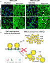
LD size decreases toward maturity in pennycress embryos. (A) Representative confocal laser‐scanning images of Arabidopsis and pennycress embryos at early developmental stages and maturity. Notice that in pennycress wild‐type plants (with LDIP) there is a noticeable shift from larger‐sized LDs in the early developmental stages to very small sized LDs at maturity, whereas in Arabidopsis embryos the LDs at maturity are only modestly smaller than those at early stages. In the mutants (without LDIP) the LDs in Arabidopsis embryos are much larger at both early development and at maturity. In pennycress mutants, the LDs at early development are somewhat larger in size compared to those in wild‐type pennycress embryos, and they become much larger at maturity without LDIP. Bar = 5 μm. (B) A hypothetical model for function of pennycress LDIP in conjunction with other LD associated proteins‐SEIPINs and LDAPs in seeds (and pollen grains). We hypothesize that pennycress LDIP may function in early embryo development in cooperation with SEIPIN, similar to Arabidopsis, and also function during maturation to reduce LD size, a feature that does not seem to be as prominent in Arabidopsis. See main text for additional details. Adapted partly from Pyc et al. (2021).

References

    1. Alameldin, H. , Izadi‐Darbandi, A. , Smith, S.A. , Balan, V. , Jones, A.D. , Ebru Orhun, G. et al. (2017) Metabolic engineering to increase the corn seed storage lipid quantity and change its compositional quality. Crop Science, 57, 1854–1864.
    1. Andrews, S. (2010) FastQC: a quality control tool for high throughput sequence data. UK: Babraham Institute.
    1. Arias, C.L. , Garcia Navarrete, L.T. , Mukundi, E. , Swanson, T. , Yang, F. , Hernandez, J. et al. (2023) Metabolic and transcriptomic study of pennycress natural variation identifies targets for oil improvement. Plant Biotechnology Journal, 21, 1887–1903. - PMC - PubMed
    1. Brocard, L. , Immel, F. , Coulon, D. , Esnay, N. , Tuphile, K. , Pascal, S. et al. (2017) Proteomic analysis of lipid droplets from Arabidopsis aging leaves brings new insight into their biogenesis and functions. Frontiers in Plant Science, 8, 894. - PMC - PubMed
    1. Cai, Y. , Goodman, J.M. , Pyc, M. , Mullen, R.T. , Dyer, J.M. & Chapman, K.D. (2015) Arabidopsis SEIPIN proteins modulate triacylglycerol accumulation and influence lipid droplet proliferation. Plant Cell, 27, 2616–2636. - PMC - PubMed

MeSH terms