. 2024 Nov 1;28(6):569-576.
doi: 10.4196/kjpp.2024.28.6.569.

Norepinephrine triggers glutamatergic long-term potentiation in hypothalamic paraventricular nucleus magnocellular neuroendocrine cells through postsynaptic β1-AR/PKA signaling pathway in vitro in rats

Affiliations

Norepinephrine triggers glutamatergic long-term potentiation in hypothalamic paraventricular nucleus magnocellular neuroendocrine cells through postsynaptic β1-AR/PKA signaling pathway in vitro in rats

Jing-Ri Jin et al. Korean J Physiol Pharmacol. .

Abstract

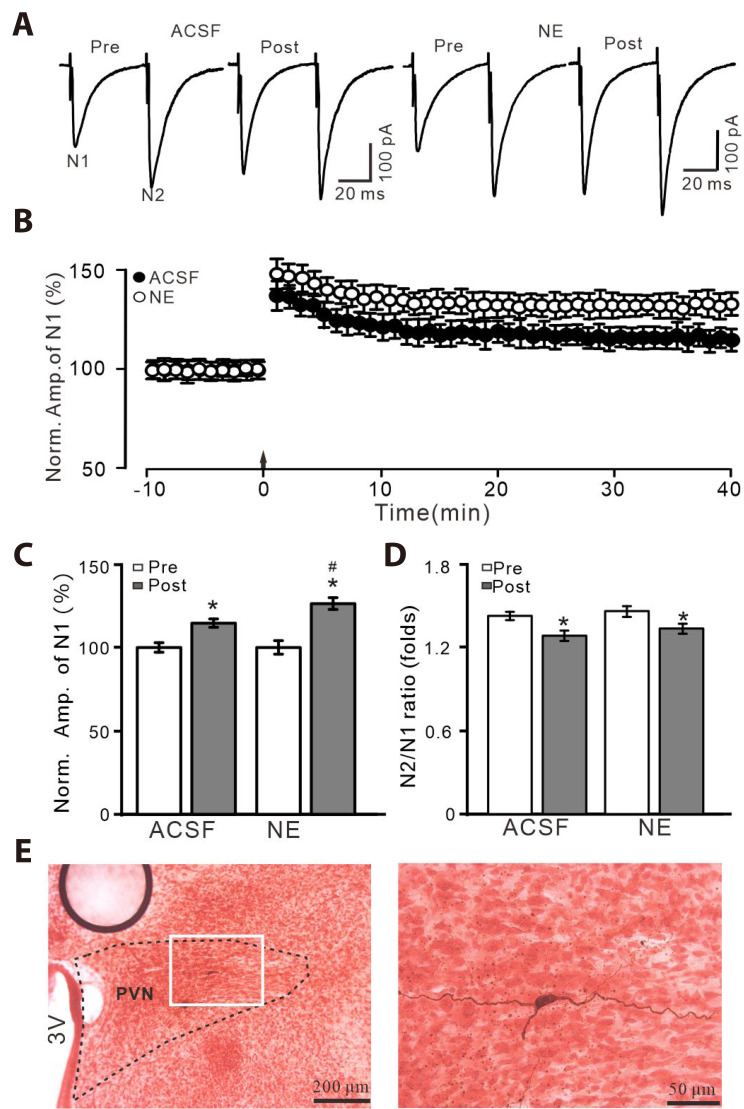
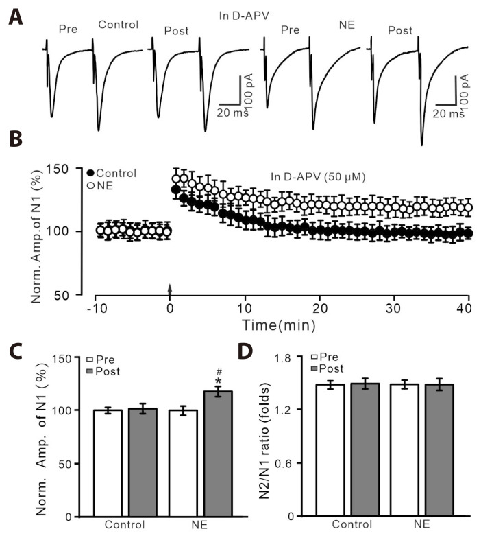
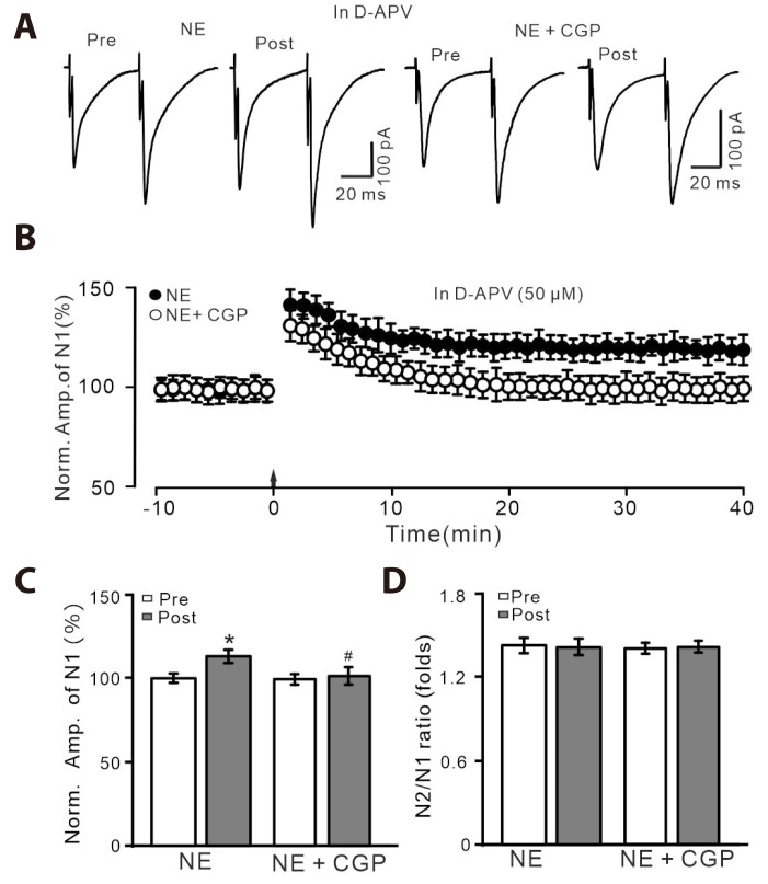
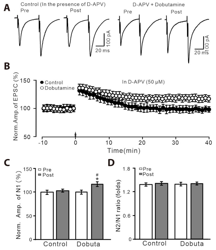
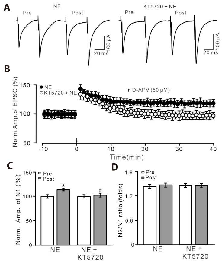
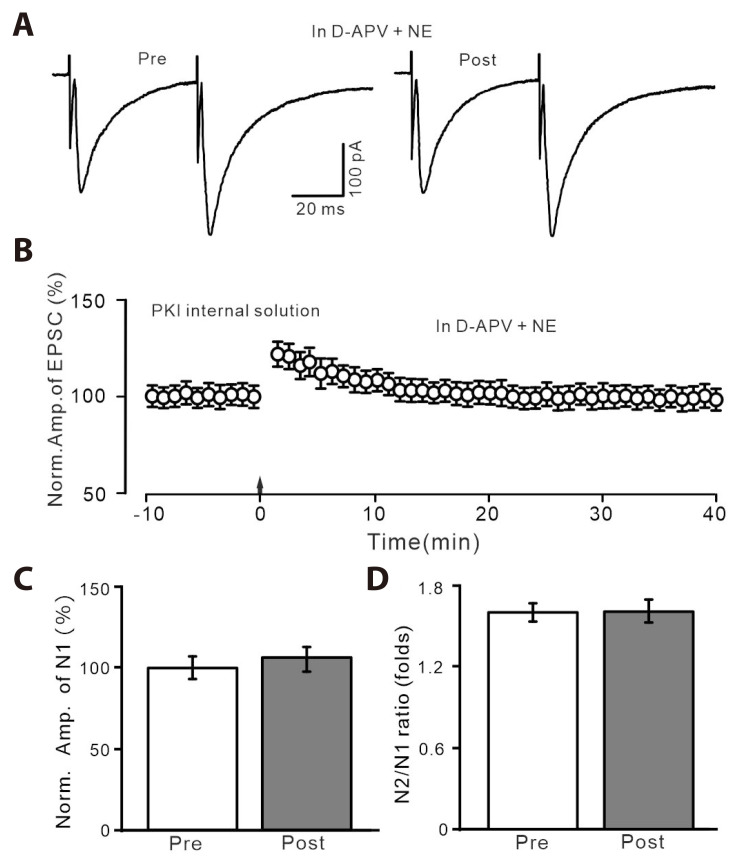
Norepinephrine (NE) modulates synaptic transmission and long-term plasticity through distinct subtype adrenergic receptor (AR)-mediated-intracellular signaling cascades. However, the role of NE modulates glutamatergic long-term potentiation (LTP) in the hypothalamic paraventricular nucleus (PVN) magnocellular neuroendocrine cells (MNCs) is unclear. We here investigate the effect of NE on high frequency stimulation (HFS)-induced glutamatergic LTP in rat hypothalamic PVN MNCs in vitro, by whole-cell patch-clamp recording, biocytin staining and pharmacological methods. Delivery of HFS induced glutamatergic LTP with a decrease in N2/N1 ratio in the PVN MNCs, which was enhanced by application of NE (100 nM). HFS-induced LTP was abolished by the blockade of N-methyl-D-aspartate receptors (NMDAR) with D-APV, but it was rescued by the application of NE. NE failed to rescue HFS-induced LTP of MNCs in the presence of a selective β1-AR antagonist, CGP 20712. However, application of β1-AR agonist, dobutamine HCl rescued HFS-induced LTP of MNCs in the absence of NMDAR activity. In the absence of NMDAR activity, NE failed to rescue HFS-induced MNC LTP when protein kinase A (PKA) was inhibited by extracellular applying KT5720 or intracellular administration of PKI. These results indicate that NE activates β1-AR and triggers HFS to induce a novel glutamatergic LTP of hypothalamic PVN NMCs via the postsynaptic PKA signaling pathway in vitro in rats.

Keywords: Adrenergic receptor; Long-term potentiation; Magnocellular neuroendocrine neuron; Paraventricular hypothalamic nucleus; Synaptic transmission.


Conflict of interest statement

CONFLICTS OF INTEREST

The authors declare no conflicts of interest.

Figures

Fig. 1
Fig. 1. NE enhanced HFS-induced glutamatergic LTP of PVN MNCs.
(A) Representative whole-cell recording traces showing paired-pulse stimulation (duration: 0.2 msec, interval: 50 msec) evoked EPSCs in the PVN MNCs before (pre), after (post) delivering HFS (100 pulses, 3 times, 10 sec interval) in treatment with ACSF (left) and NE (100 nM; right). (B) Summary of data showing the normalized amplitude of N1 before and after the delivery of HFS (arrow head) in each treatment. (C, D) Bar graphs show normalized amplitude of N1 (C) and N2/N1 ratio (D) before (pre) and after (post) the delivery of HFS. (E) Biocytin staining showing microscopic image (left, ×4) and enlarged microphotograph (right, ×40) illustrated the morphology of recorded MNC. NE, norepinephrine; HFS, high frequency stimulation; LTP, long-term potentiation; PVN, paraventricular nucleus; MNCs, magnocellular neuroendocrine cells; EPSCs, excitatory postsynaptic currents; ACSF, artificial cerebrospinal fluid. *p < 0.05 vs. pre; #p < 0.05 vs. post of control; n = 7 cells from 5 mice in each group.
Fig. 2
Fig. 2. HFS-induced LTP was abolished by an NMDAR blocker, D-APV, but it was rescued by the application of NE.
(A) In the presence of D-APV (50 µM), representative whole-cell recording traces showing paired-pulse stimulation (duration: 0.2 msec, interval: 50 msec) evoked EPSCs in PVN MNCs before (pre), after (post) delivering HFS in treatment with ACSF (control; left) and NE (100 nM; right). (B) Pooled data showing the time course of the normalized amplitude of N1 before and after delivery of HFS (arrow head) during treatment with ACSF and NE. (C, D) Bar graphs show the normalized amplitude of N1 (C) and N2/N1 ratio (D) in each group, before (pre) and after (post) delivery of HFS. HFS, high frequency stimulation; LTP, long-term potentiation; NMDAR, N-methyl-D-aspartate receptors; D-APV, D-(-)-2-Amino-5-phosphonopentanoic acid; NE, norepinephrine; EPSCs, excitatory postsynaptic currents; PVN, paraventricular nucleus; MNCs, magnocellular neuroendocrine cells; ACSF, artificial cerebrospinal fluid. *p < 0.05 vs. pre; #p < 0.05 vs. post of control; n = 7 cells from 6 mice in each group.
Fig. 3
Fig. 3. In the presence of D-APV, the NE-rescued LTP was abolished by
β1-AR antagonist. (A) In the presence of D-APV (50 µM), representative whole-cell recording traces showing paired-pulse stimulation (duration: 0.2 msec, interval: 50 msec) evoked EPSCs in PVN MNCs before (pre), after (post) delivering HFS during treatment with NE (100 nM; left) and a mixture of NE (100 nM) + CGP 20712 (1 µM). (B) In the presence of D-APV, pooled data showing the time course of normalized amplitude of N1 before and after the delivery of HFS (arrow head) during treatment with NE and a mixture of NE + CGP 20712. (C, D) Bar graphs show the normalized amplitude of N1 (C) and N2/N1 ratio (D) in each group, before (pre) and after (post) the delivery of HFS. D-APV, D-(-)-2-Amino-5-phosphonopentanoic acid; NE, norepinephrine; LTP, long-term potentiation; AR, adrenergic receptor; EPSCs, excitatory postsynaptic currents; PVN, paraventricular nucleus; MNCs, magnocellular neuroendocrine cells. *p < 0.05 vs. pre; #p < 0.05 vs. post of control; n = 8 cells from 6 mice in each group.
Fig. 4
Fig. 4. HFS-induced LTP was abolished by D-APV, but it was reversed by
β1-AR agonist. (A) In the presence of D-APV (50 µM), representative whole-cell recording traces showing paired-pulse stimulation (duration: 0.2 msec, interval: 50 msec) evoked EPSCs in PVN MNCs before (pre), after (post) delivering HFS during treatment with NE (100 nM; left) and a mixture of NE + dobutamine (1 µM; right). (B) Pooled data showing the time course of normalized amplitude of N1 before and after the delivery of HFS (arrow head) during treatment with D-APV (50 µM) and a mixture of D-APV + dobutamine. (C, D) Bar graphs show the normalized amplitude of N1 (C) and N2/N1 ratio (D) in each group, before (pre) and after (post) delivery of HFS. HFS, high frequency stimulation; LTP, long-term potentiation; D-APV, D-(-)-2-Amino-5-phosphonopentanoic acid; AR, adrenergic receptor; EPSCs, excitatory postsynaptic currents; PVN, paraventricular nucleus; MNCs, magnocellular neuroendocrine cells. *p < 0.05 vs. pre; #p < 0.05 vs. post of control; n = 7 cells from 5 mice in each group.
Fig. 5
Fig. 5. In the presence of D-APV and KT5720, NE could not rescue LTP of PVN MNCs.
(A) In the presence of D-APV (50 µM), representative whole-cell recording traces showing paired-pulse stimulation (duration: 0.2 msec, interval: 50 msec) evoked EPSCs in PVN MNCs before (pre), after (post) delivering HFS during treatment with NE (100 nM; left) and a mixture of NE (100 nM) + KT5720 (100 nM; right). (B) Pooled data showing the time course of the normalized amplitude of N1 before and after delivery of HFS (arrow head) during treatment with NE and a mixture of NE + KT5720. (C, D) Bar graphs show the normalized amplitude of N1 (C) and N2/N1 ratio (D) in each group, before (pre) and after (post) delivery of HFS. D-APV, D-(-)-2-Amino-5-phosphonopentanoic acid; NE, norepinephrine; LTP, long-term potentiation; PVN, paraventricular nucleus; MNCs, magnocellular neuroendocrine cells; EPSCs, excitatory postsynaptic currents; HFS, high frequency stimulation. *p < 0.05 vs. pre; #p < 0.05 vs. post of control; n = 9 cells from 6 mice in each group.
Fig. 6
Fig. 6. In the presence of D-APV and recording with PKI internal solution, NE could not rescue LTP of PVN MNCs.
(A) Representative current traces showing the evoked EPSCs recorded with PKI internal solution from a PVN MNCs before (pre) and after (post) delivering HFS during treatment with D-APV (50 µM) + NE (100 nM). (B) Pooled data showing the time course of normalized amplitude of N1 before and after delivery of HFS (arrow head) during treatment with D-APV + NE. (C, D) Bar graphs show the normalized amplitude of N1 (C) and N2/N1 ratio (D) before (pre) and after (post) delivery of HFS. D-APV, D-(-)-2-Amino-5-phosphonopentanoic acid; PKI, protein kinase inhibitor; NE, norepinephrine; LTP, long-term potentiation; PVN, paraventricular nucleus; MNCs, magnocellular neuroendocrine cells; EPSCs, excitatory postsynaptic currents; HFS, high frequency stimulation. *p < 0.05 vs. pre; #p < 0.05 vs. post of control; n = 6 cells from 5 mice in each group.

References

    1. Zhang BB, Jin H, Bing YH, Zhang XY, Chu CP, Li YZ, Qiu DL. A nitric oxide-dependent presynaptic LTP at glutamatergic synapses of the PVN magnocellular neurosecretory cells in vitro in rats. Front Cell Neurosci. 2019;13:283. doi: 10.3389/fncel.2019.00283. - DOI - PMC - PubMed
    1. Csáki A, Kocsis K, Halász B, Kiss J. Localization of glutamatergic/aspartatergic neurons projecting to the hypothalamic paraventricular nucleus studied by retrograde transport of [3H]D-aspartate autoradiography. Neuroscience. 2000;101:637–655. doi: 10.1016/S0306-4522(00)00411-5. - DOI - PubMed
    1. van den Pol AN, Wuarin JP, Dudek FE. Glutamate, the dominant excitatory transmitter in neuroendocrine regulation. Science. 1990;250:1276–1278. doi: 10.1126/science.1978759. - DOI - PubMed
    1. Decavel C, Van den Pol AN. GABA: a dominant neurotransmitter in the hypothalamus. J Comp Neurol. 1990;302:1019–1037. doi: 10.1002/cne.903020423. - DOI - PubMed
    1. Wuarin JP, Dudek FE. Patch-clamp analysis of spontaneous synaptic currents in supraoptic neuroendocrine cells of the rat hypothalamus. J Neurosci. 1993;13:2323–2331. doi: 10.1523/JNEUROSCI.13-06-02323.1993. - DOI - PMC - PubMed