Review
. 2024 Dec;24(12):593-609.
doi: 10.1007/s11910-024-01382-7. Epub 2024 Oct 29.

Immune Response in Traumatic Brain Injury

Affiliations
Review

Immune Response in Traumatic Brain Injury

Eder Cáceres et al. Curr Neurol Neurosci Rep. 2024 Dec.

Abstract

Purpose of review: This review aims to comprehensively examine the immune response following traumatic brain injury (TBI) and how its disruption can impact healing and recovery.

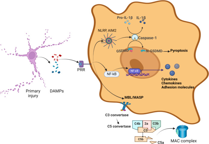
Recent findings: The immune response is now considered a key element in the pathophysiology of TBI, with consequences far beyond the acute phase after injury. A delicate equilibrium is crucial for a healthy recovery. When this equilibrium is disrupted, chronic inflammation and immune imbalance can lead to detrimental effects on survival and disability. Globally, traumatic brain injury (TBI) imposes a substantial burden in terms of both years of life lost and years lived with disability. Although its epidemiology exhibits dynamic trends over time and across regions, TBI disproportionally affects the younger populations, posing psychosocial and financial challenge for communities and families. Following the initial trauma, the primary injury is succeeded by an inflammatory response, primarily orchestrated by the innate immune system. The inflammasome plays a pivotal role during this stage, catalyzing both programmed cell death pathways and the up-regulation of inflammatory cytokines and transcription factors. These events trigger the activation and differentiation of microglia, thereby intensifying the inflammatory response to a systemic level and facilitating the migration of immune cells and edema. This inflammatory response, initially originated in the brain, is monitored by our autonomic nervous system. Through the vagus nerve and adrenergic and cholinergic receptors in various peripheral lymphoid organs and immune cells, bidirectional communication and regulation between the immune and nervous systems is established.

Keywords: Adaptive immunity; Autonomic system; Brain injury; Cholinergic pathway; Immune response; Inflammasome; Inflammation; Innate immunity; Trauma; Vagus nerve.


Conflict of interest statement

The authors declare no competing interests.

Figures

Fig. 1
Fig. 1
Innate immunity. The primary insult from external forces leads to cellular disruption and the release of membrane and cytosolic components that function as damage-associated molecular patterns (DAMPs). These DAMPs include membrane fragments, heat shock proteins (HSPs), high mobility group box 1 (HMGB1), and mitochondrial DNA, among others. DAMPs are recognized as ligands by various pattern recognition receptors (PRRs), such as toll-like receptors (TLRs), nucleotide-binding oligomerization domain-like receptors (NLRs), AIM2-like receptors (ALRs), C-type lectin receptors (CLRs), and scavenger receptors, which are linked to several intracellular pathways. ALRs and NLRs facilitate the assembly of the AIM2 and NLRP inflammasomes, which converge to activate caspase-1. Caspase-1, in turn, mediates the cleavage of pro-IL-1β into its active form, IL-1β, and gasdermin D (GSDMD) into its active form, N-GSDMD. GSDMD is an effector of the pyroptosis cell death pathway due to its ability to form pores in the cell membrane, leading to cell swelling and rupture. Additionally, IL-1β induces the expression of other cytokines such as IL-2 and IFN-γ and stimulates the activation and differentiation of T and B cells. TLRs initiate dimerization and recruit adapter proteins, such as MyD88 and TRIF, which activate signaling pathways and lead to the translocation of NF-κB. This process results in the production of cytokines, chemokines, and adhesion molecules, which facilitate the migration and infiltration of neutrophils and monocytes from the peripheral circulation. Another PRR family, CLRs—specifically mannose-binding lectin (MBL)—forms a complex with serine proteases, known as MASP (mannose-binding lectin-associated serine proteases), which activates the lectin complement pathway. This pathway leads to the formation of the membrane attack complex (MAC), which has a cytolytic effect by forming pores in the cell membrane. Acronyms: ALR: AIM2-like receptor; CLR: C-type lectin receptor; DAMP: Damage;associated molecular pattern; GSDMD: Gasdermin D; HMGB1: High mobility group box 1; HSP: Heat shock protein; MAC: Membrane attack complex; MBL: Mannose;binding lectin; MASP: Mannose;binding lectin-associated serine protease; NLR: Nucleotide-binding oligomerization domain-like receptor; PRR: Pattern recognition receptor; TLR: Toll-like receptor
Fig. 2
Fig. 2
Immune Cell Recruitment and Differentiation in Response to Brain Injury. Recruitment of T lymphocytes into the injured tissue is facilitated by the upregulation of cell adhesion molecules on vascular endothelium. These adhesion molecules and chemokines are expressed in the leptomeninges and choroid plexus. Upon arrival at the site of injury, T cells come into contact with antigen-presenting cells (APCs), such as microglia and perivascular macrophages, which present antigen peptides to T cells through Major Histocompatibility Complex (MHC) Class I and II. MHC Class I is expressed on nearly all nucleated cells and presents endogenous peptides that are recognized by cytotoxic CD8 + T cells. These T cells produce pore-forming enzymes (PFN) and granzyme B (GzmB), inducing cytolysis and apoptosis through inflammatory mediators. In contrast, the MHC Class II/antigen complex is presented by APCs to naïve CD4 + T lymphocytes via the T cell receptor (TCR). MHC Class II antigen, in conjunction with other costimulatory molecules like CD80 and CD86, initiates a network of internal signaling that leads to the differentiation of naïve CD4 + T lymphocytes into five major types of CD4 + T helper (Th) cells: Th1, Th2, Th17, T regulatory (Treg), and T follicular helper (Tfh) cells. Th1 cells play a pro-inflammatory role by producing IL-2, TNF-α, and IFN-γ. Th17 cells also amplify inflammation through the production of IL-17, IL-22, and IL-23. Th2 cells produce anti-inflammatory or modulatory cytokines, including IL-4, IL-5, IL-10, and IL-13, and contribute to tissue repair by activating macrophages that induce fibroblast proliferation and the removal of extracellular matrix and apoptotic cells. Treg cells promote immune tolerance and attenuate T cell brain infiltration by expressing IL-10 and TGF-β. Tfh cells migrate to lymphoid tissue via the meningeal lymphatic system, initially reaching the cervical lymph nodes, where they present the MHC Class II/antigen complex to the B cell receptor (BCR) of B cells, thereby inducing the development of antigen-specific B cell immunity. T cells must display the CD40 ligand (CD40L), which binds to the CD40 receptor on the B cell surface as a costimulatory signal, leading to the maturation and activation of B cells. Acronyms: APC: Antigen-presenting cell; BCR: B cell receptor; CD40L: CD40 ligand; GzmB: Granzyme B; IFN-γ: Interferon-gamma; IL: Interleukin; MHC: Major Histocompatibility complex; PFN: Perforin; TCR: T cell receptor; Th: CD4 + T helper cell; Tfh: T follicular helper cell; Treg: T regulatory cell; TNF-α: Tumor necrosis factor-α; TGF-β: Transforming growth factor-β
Fig. 3
Fig. 3
The Neuroimmune Reflex in Response to Trauma. Activation of the vagus nerve by inflammatory cytokines following trauma triggers a reflex response involving the celiac ganglion. This ganglion, which is sympathetically connected to the spleen, stimulates the release of norepinephrine. Norepinephrine activates β2-adrenergic receptors on splenic CD4 + T lymphocytes, which in turn produce acetylcholine (ACh). ACh further stimulates splenic leukocytes, such as macrophages and dendritic cells, via the α7 nicotinic acetylcholine receptor (α7nAChR), resulting in a reduction of pro-inflammatory cytokines like TNF-α. Acronyms: ACh: Acetylcholine; α7nAChR: α7 Nicotinic Acetylcholine Receptor; TNF-α: Tumor Necrosis Factor-α

References

    1. Taylor CA, Bell JM, Breiding MJ, Xu L. Traumatic Brain injury-related emergency department visits, hospitalizations, and deaths - United States, 2007 and 2013. MMWR Surveill Summ. 2017;66(9):1–16. 10.15585/mmwr.ss6609a1. - PMC - PubMed
    1. Steyerberg EW, Wiegers E, Sewalt C, Buki A, Citerio G, De Keyser V, Ercole A, Kunzmann K, Lanyon L, Lecky F, Lingsma H, Manley G, Nelson D, Peul W, Stocchetti N, von Steinbüchel N, Vande Vyvere T, Verheyden J, Wilson L, Maas AIR, Menon DK, CENTER-TBI Participants and Investigators. Case-mix, care pathways, and outcomes in patients with traumatic brain injury in CENTER-TBI: a European prospective, multicentre, longitudinal, cohort study. Lancet Neurol. 2019;18(10):923–34. 10.1016/S1474-4422(19)30232-7. - PubMed
    1. Meyfroidt G, Bouzat P, Casaer MP, Chesnut R, Hamada SR, Helbok R, Hutchinson P, Maas AIR, Manley G, Menon DK, Newcombe VFJ, Oddo M, Robba C, Shutter L, Smith M, Steyerberg EW, Stocchetti N, Taccone FS, Wilson L, Zanier ER, Citerio G. Management of moderate to severe traumatic brain injury: an update for the intensivist. Intensive Care Med. 2022;48(6):649–66. 10.1007/s00134-022-06702-4. (Erratum in: Intensive Care Med. 2022 Jul;48(7):989-991. 10.1007/s00134-022-06759-1). - PubMed
    1. Shanahan R, Avsar P, Watson C, Moore Z, Patton D, McEvoy NL, Curley G, O’Connor T. The impact of brain tissue oxygenation monitoring on the Glasgow Outcome Scale/Glasgow Outcome Scale Extended in patients with moderate to severe traumatic brain injury: A systematic review. Nurs Crit Care. 2023. 10.1111/nicc.12973. - PubMed
    1. McDonald BZ, Tarudji AW, Zhang H, Ryu S, Eskridge KM, Kievit FM. Traumatic brain injury heterogeneity affects cell death and autophagy. Exp Brain Res. 2024;242(7):1645–58. 10.1007/s00221-024-06856-1. - PubMed