. 2024 Oct 28;14(1):25818.
doi: 10.1038/s41598-024-75985-y.

Cationized extracellular vesicles for gene delivery

Affiliations

Cationized extracellular vesicles for gene delivery

Natalia L Klyachko et al. Sci Rep. .

Abstract

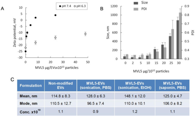
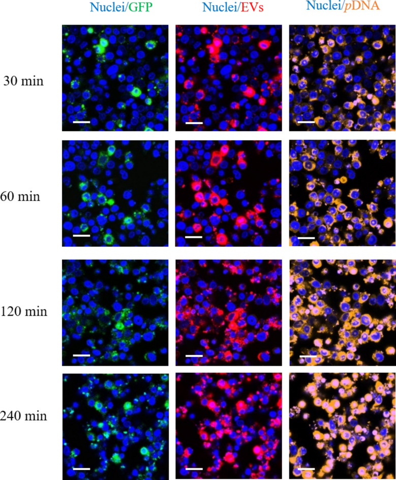
Last decade, extracellular vesicles (EVs) attracted a lot of attention as potent versatile drug delivery vehicles. We reported earlier the development of EV-based delivery systems for therapeutic proteins and small molecule chemotherapeutics. In this work, we first time engineered EVs with multivalent cationic lipids for the delivery of nucleic acids. Stable, small size cationized EVs were loaded with plasmid DNA (pDNA), or mRNA, or siRNA. Nucleic acid loaded EVs were efficiently taken up by target cells as demonstrated by confocal microscopy and delivered their cargo to the nuclei in triple negative breast cancer (TNBC) cells and macrophages. Efficient transfection was achieved by engineered cationized EVs formulations of pDNA- and mRNA in vitro. Furthermore, siRNA loaded into cationized EVs showed significant knockdown of the reporter gene in Luc-expressing cells. Overall, multivalent cationized EVs represent a promising strategy for gene delivery.

Keywords: Cancer; EVs; Gene delivery; Multivalent cationic lipid; Transfection.


Conflict of interest statement

The authors declare no competing interests.

Figures

Fig. 1
Fig. 1
Overall scheme for preparation of EV-nucleic acid nanoformulation.
Fig. 2
Fig. 2
Characterization of the MVL5 cationized EVs (MVL5-EVS): (A) zeta-potential, (B) particle size and PDI by DLS and (C) particle size and concentration by NTA depending on the method of preparation of MLV5-EVs. The MLV5-EVs were prepared as indicated using EVs dispersed (A) in 10 mM phosphate buffer at pH 6.3 or (A, B) pH 7.4; and (C) in PBS or 50% PBS/EtOH solution with sonication or saponin permeabilization of EVs membranes. Addition of cationic lipid resulted in the increase of the (A) zeta-potential of EVs at both pH, and (B) size and PDI of the particles. The size and concentration of MLV5-EVs (2 μg/1010 particles) manufactured at various conditions were measured by NTA (C). Non-incorporated MLV5 was removed by size exclusion chromatography (A, B) or PEG precipitation (C) before the measurements.
Fig. 3
Fig. 3
Detection of pDNA compaction by EtBr displacement assay. The pDNA was mixed with EtBr and then titrated with MVL5-EVs. Sample fluorescence was determined after subtracting the baseline fluorescence of EtBr in the absence of pDNA. Data are presented as the ratio of fluorescence of EtBr (1 µg/mL) with pDNA (12.8 µg/µL) in the EVs with and without MVL5. Intercalation of EtBr into pDNA is accompanied with the increase of fluorescence quantum yield of the dye. Addition of the cationic lipid MVL5 results in the quenching of the fluorescence due to the displacement of EtBr by the MVL5.
Fig. 4
Fig. 4
Intracellular distribution of cationized EVs-pDNA. YOYO-labeled pDNA (here green) was loaded into EVs and purified by PEG precipitation. Cationized EVs-YOYO-pDNA (1 × 1011 particles/mL) was added to RAW 264.7 macrophages for various time points. Following incubation, the cells were washed with PBS, and nuclei were stained with Hoechst (here red). Significant co-localization of pDNA and nuclei staining was recorded over 240 min, indicating that EVs delivered pDNA to the nuclei. pDNA (1.6 µg/µL); EVs (1 × 1011 particles); cationic lipid, PEI (27 mg/mL) PEI was incorporated into EVs by transient permeabilization with saponin as described in Materials and Methods. The bar: 20 μm.
Fig. 5
Fig. 5
Accumulation of MVL5-EVs/CY5-siRNA in cancer cells. MDA-MB-231 cells (1 × 104 cells/well) were incubated with: (A) CY5-siRNA alone (1 nmol/well, red), (B) mixture of CY5-siRNA and non-modified EVs, (C) MVL5 lipid and siRNA, (D) siRNA loaded into MVL5-EVs (10 µg MVL5/1010 particles) by sonication, and (E) siRNA loaded into MVL5-EVs (the same number of particles) by saponin. The nuclei were stained with Hoechst (blue). (F) The mean fluorescence intensity of CY5-siRNA in the cells was determined. After MVL5 incorporation into EVs by sonication or saponin permeabilization, MVL5-EVs were purified by column chromatography, mixed with CY5-siRNA, and then purified again. The bar: 20 μm.
Fig. 6
Fig. 6
Transfection of IC21 macrophages with MVL5-EVs-based formulations of pDNA. (A) MVL5-EVs (20 µg MVL5/1011particles) were prepared via MVL5 incorporation into EVs by sonication at different temperatures (PEG purification after MVL5 incorporation), and then loaded with luciferase pDNA (6 µg /1011particles). IC21 macrophages (1 × 105 cells/well) were supplemented with MVL5-EVs formulations of pDNA (1 × 1010 particles/1 µg pDNA/well) and incubated for 4 h at 37 °C. (B) MVL5-EVs were prepared via incorporation of different amounts of MVL5 (0.5–50 µg MVL5/1011particles) into EVs by sonication in the presence or absence of EtOH (50%) and added to IC21 macrophages (2 × 104 cells/well). Free pDNA (1 µg/well), or mixture of EVs and pDNA (1010particles/1 µg pDNA/well), or mixture of common transfection agent, GenePorter (GP3K, according to manufacturer’s protocol), with pDNA (1 µg/well) at the same concentration were used as controls. Then, the cells were washed with PBS and cultured in full media for 24 h, and the luminescence of lysed cells in the presence of luciferin-ATP was measured. White bars – samples without EVs. (A) Bell shaped curve was observed for transfection efficacy with maximum at 42 °C used for cationized EVs preparation. (B) Transfection efficacy increased with the increases of MVL5 incorporated into EVs nanocarriers. Little, if any, luminescence was detected in cells exposed to naked pDNA, or mixture pDNA with non-cationized EVs. As expected, significant transfection was observed in the cells supplemented mixture of free pDNA and the cationic lipid, or free pDNA and GP3K.
Fig. 7
Fig. 7
Accumulation of cationized EVs loaded with GFP-pDNA, and the expression of the encoded protein in Raw 264.7 macrophages. Cationized EVs were labeled with fluorescent dye, DID (red) and GFP-encoding pDNA was labeled with nucleic acid fluorescent dye, TOTO (orange, ex/em 514/533). Raw 264.7 macrophages (5 × 104 cells/well) were supplemented with cationized EVs loaded with GFP-pDNA (1010 particles/1µg pDNA/well) for various times, and intracellular distribution of all components of the formulation, as well as the expression of the encoded protein, GFP, was examined by confocal microscopy. The nuclei were stained with Hoechst (blue). Considerable amount of cationized EVs, as well as TOTO-pDNA was detected in Raw 264.7 macrophages over 240 min. The expression of GFP was increased at later time points (120–240 min). Cationized EVs were obtained by EVs supplemented with PEI by saponin permeabilization, followed by PEG purification. The bar: 20 μm.
Fig. 8
Fig. 8
Transfection IC21 macrophages with MVL5-EVs-luc-mRNA. MVL5-EVs (20 µg MVL5/1011particles) were prepared via MVL5 incorporation into EVs by sonication at 42 °C with PEG purification after MVL5 incorporation, and then loaded with luciferase mRNA (1.3 µg /1011 particles). IC21 macrophages (1 × 105 cells/well) were supplemented with MVL5-EVs-Luc-mRNA (3 µg/1011 particles/well) loaded with different amount of mRNA (0.4–3 µg) and incubated for 4 h at 37 °C. Naked mRNA (maximum amount, 3 µg/well) with or without GP3K was used as a control. Then, the cells were washed with PBS and cultured in full media for 24 h, and the luminescence of lysed cells in the presence of luciferin-ATP was measured. Cationized EVs provided efficient transfection in vitro. MVL5-EVs were obtained by lipid incorporation using sonication in water bath followed by PEG precipitation.
Fig. 9
Fig. 9
Knockdown of Luc by MVL5-EVs-siRNA in Luc-MDA-MB-231 cancer cells. (A) MVL5-EVs (12 µg MVL5/1010particles) were prepared via MVL5 incorporation into EVs by sonication at 420C with PEG purification after MVL5 incorporation, and then 65 µL of MVL5-EVs were loaded with GL2 + GL3 siRNA (0.6 µM /1010 particles). Previously transfected MDA-MB-231 cells (Luc-MDA-MB-231) (1 × 104 cells/well) were supplemented with MVL5-EVs loaded with GL2 + GL3 siRNA (1.3 µg MVL5/109 particles/40 nM siRNA/well) and incubated for 4 h at 37 °C. Then, cells were washed and cultured in complete media for 24 h. Free siRNA, or mixture siRNA with EVs, or MVL5-EVs without siRNA, or mixture of siRNA and MVL5, or scrambled siRNA (control #1) at same concentrations were used as controls. Free media was also used as control. The luminescence was measured in cell lysates. Significant knockdown of Luc was recorded in the cells incubated by MVL5-EVs-siRNA. *p < 0.0001 compared to media; #p < 0.001 compared to siRNA mixed with MVL5. (B) MVL5-EVs (12 µg MVL5/1010particles) were prepared via MVL5 incorporation into EVs by sonication at 420C with PEG purification after MVL5 incorporation, and then 65 µL of MVL5-EVs were loaded with GL2 + GL3 siRNA at different concentrations of siRNA (0.6 µM, 1.23 µM, or 2.46 µM). Luc-MDA-MB-231 cells (1 × 104 cells/well) were supplemented with different concentrations of MVL5-EVs loaded with GL2 + GL3 siRNA (up to 80 nM siRNA) and incubated for 4 h at 370C. Then, cells were washed and cultured in complete media for 24 h. Increases in siRNA amount in MVL5-EVs formulation resulted in the greater knockdown of the target protein ***p < 0.00005; ****p < 0.000005 compared to Luc-MDA-MB-231 with scramble siRNA, the first bar on the graph.

Similar articles

Cited by

References

    1. Saraiva, C. et al. Nanoparticle-mediated brain drug delivery: overcoming blood–brain barrier to treat neurodegenerative diseases. J. Control Release. 235, 34–47 (2016). - PubMed
    1. Zhang, Q. et al. Stimuli-responsive gene delivery nanocarriers for cancer therapy. Nano-Micro Lett. 15, 44 (2023). - PMC - PubMed
    1. Movahedi, F., Hu, R. G., Becker, D. L. & Xu, C. Stimuli-responsive liposomes for the delivery of nucleic acid therapeutics. Nanomed. Nanotechnol. Biol. Med. 11, 1575–1584 (2015). - PubMed
    1. Dirisala, A. et al. Effective mRNA protection by poly (l-ornithine) synergizes with endosomal escape functionality of a charge‐conversion polymer toward maximizing mRNA introduction efficiency. Macromol. Rapid Commun. 43, 2100754 (2022). - PubMed
    1. Lopukhov, A. V. et al. Mannosylated cationic copolymers for gene delivery to macrophages. Macromol. Biosci. 21, 2000371 (2021). - PMC - PubMed

LinkOut - more resources