. 2024 Oct;30(10):e70093.
doi: 10.1111/cns.70093.

Role of Hippocampal Glutamatergic Synaptic Alterations in Sevoflurane-Induced Cognitive Dysfunction in Aged Mice

Affiliations

Role of Hippocampal Glutamatergic Synaptic Alterations in Sevoflurane-Induced Cognitive Dysfunction in Aged Mice

Yixuan Niu et al. CNS Neurosci Ther. 2024 Oct.

Abstract

Aims: Perioperative neurocognitive disorders (PND), including postoperative delirium (POD) and postoperative cognitive dysfunction (POCD), are common following anesthesia and surgery in older patients and significantly increase morbidity and mortality. However, the underlying mechanism of PND is unclear. Our study aims to analyze the differentially expressed genes (DEGs) in excitatory neurons and investigate the role of hippocampal glutamatergic synaptic alterations in sevoflurane-induced cognitive dysfunction in aged mice.

Methods: We performed single-nucleus RNA sequencing (snRNA-seq) technology to examine the alterations of excitatory neurons in hippocampus induced by sevoflurane in aged mice. Gene Ontology (GO) and Kyoto Encyclopedia of Genes and Genomes (KEGG) analysis of DEGs were performed in excitatory neurons. At last, immunofluorescence staining was used to validate sevoflurane-induced alternation of glutamatergic synapses in the hippocampus of aged mice.

Results: This study demonstrates that DEGs in excitatory neurons are associated with reduction of glutamatergic synapses and cognitive dysfunction. After immunofluorescence staining validation, we also confirmed that sevoflurane anesthesia decreased the density of glutamatergic synapses in the hippocampus of aged mice.

Conclusions: Our findings demonstrated a key role of hippocampal glutamatergic synaptic alterations in sevoflurane-induced cognitive dysfunction in aged mice.

Keywords: PND; aging; anesthesia; excitatory neuron; glutamatergic synapse; sevoflurane.


Conflict of interest statement

The authors declare no conflicts of interest.

Figures

FIGURE 1
FIGURE 1
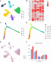
Hippocampus single‐nucleus transcriptome of aged mice. (A) Schematic graph showing the experimental design and general workflow of snRNA‐seq, including three key steps. (1) Three mice were in the control group (Ctrl). Three mice were in the sevoflurane group (SEV). (2) Hippocampus was dissected from mouse and harvested according to the Allen Brain Atlas. Each sample was used to obtain single‐nucleus suspensions. (3) Nuclei isolation was performed and snRNA‐seq was finally measured and analyzed. (B) Different snRNA‐seq markers representing different cell types, including excitatory neurons (Ex), astrocytes (Ast), endothelial cells (EC), motor neuron, microglia (Mic), interneurons, oligodendrocytes (OL), oligodendrocyte precursor cells (OPC), Temperature‐sensitive POA neurons of mouse. (C, D) UMAP representation of diverse cell types of mice, were identified by the specific snRNA‐seq marker and colored in cluster (C) or group (D).
FIGURE 2
FIGURE 2
Excitatory neuron cluster taxonomy of mice's hippocampus. (A) UMAP representation of excitatory neurons colored by cluster. (B) Heatmap of 20 snRNA‐seq marker genes in excitatory neurons of mouse. (C) The scatter plot shows the distribution of five distinct excitatory neuron clusters. Each point represents a single cell, colored by its assigned subtype. (D) Cells are colored based on their calculated stemness score, with a gradient ranging from high stemness (red) to low stemness (blue). (E) UMAP representation of excitatory neurons colored by group (red, SEV; blue, Ctrl). (F) Bar plot showing the differences in mouse excitatory neuron cluster between group SEV and Ctrl. The number of excitatory neurons following Ctrl are close to the number of excitatory neurons following SEV in each cluster of mice.
FIGURE 3
FIGURE 3
Sevoflurane anesthesia caused DEGs in mouse. (A) Heatmaps representation of 20 DEGs in excitatory neuron clusters of mice's hippocampus after sevoflurane exposure following Ctrl versus SEV. Demonstration of the heatmap indicates that sevoflurane anesthesia caused diverse changes in mouse. (B) Volcano plots representation of DEGs in excitatory neuron of mouse after sevoflurane exposure. In the volcano plot, p < 0.05 was set as the cut‐off criterion of significant difference. Red tags representation of the up‐regulated significant DEGs and green tags representation of the downregulated significant DEGs. (C) Bar graph showing the number of DEGs in excitatory neurons clusters of mice after sevoflurane exposures between Ctrl and SEV group. Red bars illustrating the number of up‐regulated genes and green bars representing the number of downregulated genes.
FIGURE 4
FIGURE 4
GO and KEGG pathway terms of DEGs in mice's hippocampus after sevoflurane anesthesia. (A) GO enrichment of DEGs in excitatory neuron of mouse, illustrating the biological functions of mouse. Bars colored by red demonstrating the GO enrichment of up‐regulated DEGs and green demonstrating the GO enrichment of downregulated DEGs. (B) Bar plots showing the KEGG enrichment in excitatory neuron of mouse. Red bars illustrating the enriched KEGG pathways of up‐regulated DEGs and green bars representing the enriched KEGG pathways of downregulated DEGs. Significant enriched biological process (gene ontology) (C) and cellular component (gene ontology) (D) terms of the DEGs (p < 0.05, FC > 1.2 or FC < 0.8) in excitatory neurons of mice's hippocampus after sevoflurane anesthesia.
FIGURE 5
FIGURE 5
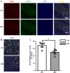
Sevoflurane downregulates the levels of PSD95 and vGLUT1 in the hippocampus of aged mice. (A, B) Glutamatergic synapses were stained by immunofluorescence (PSD95‐red, vGLUT1‐green, DAPI‐blue, merged image‐yellow) in the hippocampus of aged mice. (C) Bar plot of vGLUT1/PSD95 puncta number per 100 μm. Data are expressed as the mean ± SEM (one‐way ANOVA followed by Bonferroni's post hoc test, n = 6 per group). *p < 0.05 versus he Ctrl group.

References

    1. Evered L., Silbert B., Knopman D. S., et al., “Recommendations for the Nomenclature of Cognitive Change Associated With Anaesthesia and Surgery‐2018,” British Journal of Anaesthesia 121, no. 5 (2018): 1005–1012, 10.1016/j.bja.2017.11.087. - DOI - PMC - PubMed
    1. Luo A., Yan J., Tang X., Zhao Y., Zhou B., and Li S., “Postoperative Cognitive Dysfunction in the Aged: The Collision of Neuroinflammaging With Perioperative Neuroinflammation,” Inflammopharmacology 27, no. 1 (2019): 27–37, 10.1007/s10787-018-00559-0. - DOI - PubMed
    1. Pan C., Zhang H., Zhang L., et al., “Surgery‐Induced Gut Microbial Dysbiosis Promotes Cognitive Impairment via Regulation of Intestinal Function and the Metabolite Palmitic Amide,” Microbiome 11, no. 1 (2023): 248, 10.1186/s40168-023-01689-6. - DOI - PMC - PubMed
    1. Liu B., Huang D., Guo Y., et al., “Recent Advances and Perspectives of Postoperative Neurological Disorders in the Elderly Surgical Patients,” CNS Neuroscience & Therapeutics 28, no. 4 (2022): 470–483, 10.1111/cns.13763. - DOI - PMC - PubMed
    1. Wang D. X., Dong Z. J., Deng S. X., et al., “GDF11 Slows Excitatory Neuronal Senescence and Brain Ageing by Repressing P21,” Nature Communications 14, no. 1 (2023): 7476, 10.1038/s41467-023-43292-1. - DOI - PMC - PubMed

Publication types

MeSH terms

Associated data

LinkOut - more resources