. 2024 Oct 28;22(1):665.
doi: 10.1186/s12951-024-02941-3.

Magnetic targeting enhances the neuroprotective function of human mesenchymal stem cell-derived iron oxide exosomes by delivering miR-1228-5p

Affiliations

Magnetic targeting enhances the neuroprotective function of human mesenchymal stem cell-derived iron oxide exosomes by delivering miR-1228-5p

Wei-Jia Hu et al. J Nanobiotechnology. .

Abstract

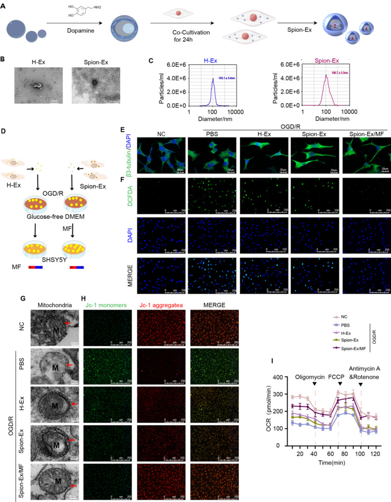
Background: Treating mitochondrial dysfunction is a promising approach for the treatment of post-stroke cognitive impairment (PSCI). HuMSC-derived exosomes (H-Ex) have shown powerful therapeutic effects in improving mitochondrial function, but the specific effects are unclear and its brain tissue targeting needs to be further optimized.

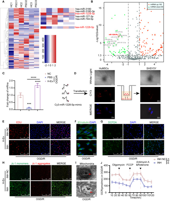
Results: In this study, we found that H-Ex can improve mitochondrial dysfunction of neurons and significantly enhance the cognitive behavior performance of MCAO mice in OGD/R-induced SHSY5Y cells and MCAO mouse models. Based on this, we have developed an exosome delivery system loaded with superparamagnetic iron oxide nanoparticles (Spion-Ex) that can effectively penetrate the blood-brain barrier (BBB). The research results showed that under magnetic attraction, Spion-Ex can more effectively target the brain tissue and significantly improve mitochondrial function of neurons after stroke. Meanwhile, we further confirmed that miR-1228-5p is a key factor for H-Ex to improve mitochondrial function and cognitive behavior both in vivo and in vitro. The specific mechanism is that the increase of miR-1228-5p mediated by H-Ex can inhibit the expression of TRAF6 and activate the TRAF6-NADPH oxidase 1 (NOX1) pathway, thereby exerting protective effects against oxidative damage. More importantly, we found that under magnetic attraction, Spion-Ex exhibited excellent cognitive improvement effects by delivering miR-1228-5p.

Conclusions: Our research found that H-Ex has a good therapeutic effect on PSCI by increasing the expression of miR-1228-5p in PSCI, while H-Ex loaded with Spion-Ex exhibited more excellent effects on improving mitochondrial function and cognitive impairment under magnetic attraction, which can be used as a novel strategy for the treatment of PSCI.

Keywords: Cognitive impairment; Exosomes; Oxidative stress; Superparamagnetic iron oxide nanoparticles.


Conflict of interest statement

The authors declare no competing interests.

Figures

Fig. 1
Fig. 1
Spion-Ex/MF alleviated oxidative damage and mitochondrial dysfunction in SHSY5Y cells after OGD/R. (A). Schematic illustration of preparation of Spion-Ex. (BC). TEM imaging (B) and NTA of particle size (C) of H-Ex and Spion-Ex. Scale bar = 200 nm (D). Co-culture flow chart. (E). Total neurite length was imaged by β3-tubulin staining. Scale bar = 20 μm, n = 3 independent experiments. (F). Intracellular DCFDA staining. Scale bar = 250 μm, n = 3 independent experiments. Green: DCFDA staining. Blue: DAPI-labeled nucleus. G. Mitochondrial morphology under TEM. Scale bar = 200 nm, n = 3 independent experiments. H. JC-1 staining was used to detect the changes of mitochondrial membrane potential. Scale bar = 250 μm, n = 3 independent experiments, Red: JC-1 aggregates. Green: JC-1 monomers. I. The analysis of OCR. n = 3 independent experiments. Spion-Ex/MF: Spion-Ex under magnetic field
Fig. 2
Fig. 2
Spion-Ex/MF safely improved cognitive and memory function in stroke. (A) Schematic illustration of preparation of Spion-Ex. (B) Representative track traces of each respective group and time spent in the central area in the open field test, n = 10 independent experiments. (C) The representative swim path showing sample paths of mice from training trials on day 7 and crossing platform times in the target quadrant in the MWM, n = 10 independent experiments. (D) Representative traces from each group in the novel object recognition (NOR) and preference of different groups of mice for new objects on the testing day of the NOR experiment, n = 10 independent experiments. *P < 0.05,**P < 0.01,***P < 0.001,****P < 0.0001, Spion-Ex/MF: Spion-Ex under magnetic field
Fig. 3
Fig. 3
Spion-Ex/MF mitigated oxidative damage to neurons in vivo. (A) Representative pictures of ROS production. Scale bar = 250 μm. (B) Image representing colocalization of NeuN (green) and Tunel (red) in dentate gyrus of hippocampus brain section. (CD). TEM images showed mitophagy (C) and (D) synaptic structures in hippocampal neuron. Scale bar = 200 nm. **P < 0.01,****P < 0.0001, Spion-Ex/MF: Spion-Ex under magnetic field
Fig. 4
Fig. 4
Spion-Ex/MF effectively increased the targeted delivery of HuMSC-derived exosomes to injured neurons. (A) Experimental design and Prussian blue reaction (B) Image of H-Ex uptake in mouse hippocampus. (C) Mice were followed by injected in H-Ex、Spion-Ex and Spion-Ex/MF, administered via tail vein injection. Coronal sections of T2-weighted images showed the labeled Spion-Ex as a hypointense area (arrowhead). (D and E). Noninvasive NIRF imaging of Dir-labeled H-Ex in mice after intravenous injection with H-Ex and Spion-Ex. Spion-Ex/MF: Spion-Ex under magnetic field
Fig. 5
Fig. 5
miR-1228-5p ameliorated oxidative stress and cognitive impairment induced by ischaemic stroke. (A) Partial heatmap of differentially expressed serum miRNAs between PSCI patients and HC subjects. (B) The volcano maps. (C) Screening purpose miRNA. (D) MSC transfected with the CY3-miR-1228-5p mimic (red fluorescence) were plated in the upper chamber and co-incubated with SHSY5Y in the lower chamber in a coculture system with a 0.4 μm pore membrane. Red fluorescence was detected in the MSC recipient cells. (E) The proliferation. Scale bar = 250 μm, n = 3 independent experiments. (F) Total neurite length. (G) Intracellular DCFDA staining. Scale bar = 250 μm, n = 3 independent experiments. Green: DCFH-DA staining. Blue: DAPI-labeled nucleus. (H) Representative JC-1 staining pictures. Scale bar = 250 μm. n = 3 independent experiments. Red: JC-1 aggregates. Green: JC-1 monomers. (I) TEM images of mitochondrial ultrastructure. Scale bar = 200 nm. n = 3 independent experiments. (J) The analysis of OCR. n = 3 independent experiments. **P < 0.01,****P < 0.0001
Fig. 6
Fig. 6
Regulation of the oxidative stress pathway by miR-1228-5p by targeting TRAF6/NOX1. (A and B) Dual luciferase assays of miR-1228-5p targeting effects on TRAF6 3’-UTR. Data are presented as means ± SEM, n = 3 independent experiments. (C and D) The protein expression levels of TRAF6 and quantified. β-actin was used as a loading control. (E and F)The level of TRAF6 (E) and NOX1(F). Data are presented as means ± SEM, n = 3. (G). The proliferation. Scale bar = 50 μm, n = 3 independent experiments. (H). Total neurite length. (I). Intracellular ROS production. Scale bar = 250 μm, n = 3 independent experiments. (J). Representative JC-1 staining pictures. Scale bar = 250 μm, n = 3 independent experiments. (K). TEM images of mitochondrial ultrastructure. Scale bar = 250 μm, n = 3 independent experiments. (L). The analysis of OCR. n = 3 independent experiments. *P < 0.05,**P < 0.01,***P < 0.001, Spion-Ex/MF: Spion-Ex under magnetic field
Fig. 7
Fig. 7
Spion-Ex-embedded miR-1228-5p protects against memory and oxidative impairment in vivo. (A) The level of miR-1228-5p in the hippocampus. (B) Representative images of immunohistochemistry results for TRAF6 protein. (C) The protein expression levels of TRAF6 (B) and NOX1 (C) and quantified. (D) Neurological deficit scores, n = 10 independent experiments. (E) Representative track traces in the OFT. (F) Quantitative analysis of total distance in the OFT, n = 10 independent experiments. (GH). Time spent (G) and distance (H) in the central area in the OFT, n = 10 independent experiments. (I). Traces on the probe trial on day 7 in the MWM. (JL). The time spent (J), crossing platform (K) and latencies (L) times in the target quadrant in the MWM, n = 10 independent experiments. (M). The preference of different groups of mice for new objects on the training day of the NOR experiment, n = 10 independent experiments. (N). The preference of different groups of mice for new objects on the testing day of the NOR experiment, n = 10 independent experiments. (O). Representative traces in the NOR. (PQ). Representative pictures of ROS production (P) and TUNEL staining (Q) in the hippocampus. Scale bar = 50 μm. (RS). TEM images showed mitophagy and synaptic structures in hippocampal neuron. Scale bar = 200 nm. *P < 0.05,**P < 0.01,***P < 0.001,****P < 0.0001,Spion-Ex: exosome delivery system loaded with superparamagnetic iron oxide nanoparticles
Fig. 8
Fig. 8
Abnormal expression of miR-1228-5p in PSCI patients. (A) The MRI representative image of patients with PSCI. (B) The miR-1228-5p levels in serum. (C) The expression of TRAF6 and NOX1. (DE). The MRS of hippocampus in PSCI patients. (F). Positive correlation between serum miR-1228-5p levels and MoCA scores in PSCI patients (n = 40) (Pearson analysis). (G). The proposed model illustrating the property of Spion-Ex/MF. ***P < 0.001,****P < 0.0001, PSCI: Post-stroke cognitive impairment

References

    1. Chi N-F, Hu H-H, Chan L, Wang C-Y, Chao S-P, Huang L-K, Ku H-L. And Hu C-J. Impaired cerebral autoregulation is associated with poststroke cognitive impairment. Ann Clin Transl Neurol. 2020;7:1092–102. - PMC - PubMed
    1. Mijajlović MD, Pavlović A, Brainin M, Heiss W-D, Quinn TJ, Ihle-Hansen HB, Hermann DM, Assayag EB, Richard E, Thiel A, Kliper E, Shin Y-I, Kim Y-H, Choi S, Jung S, Lee Y-B, Sinanović O, Levine DA, Schlesinger I, Mead G, Milošević V, Leys D, Hagberg G, Ursin MH, Teuschl Y, Prokopenko S, Mozheyko E, Bezdenezhnykh A, Matz K, Aleksić V, Muresanu D. Korczyn AD, Bornstein NM. Post-stroke dementia - a comprehensive review. BMC Med. 2017;15:11. - PMC - PubMed
    1. Fan X, Shen W, Wang L, Zhang Y. Efficacy and safety of DL-3-n-butylphthalide in the treatment of poststroke cognitive impairment: a systematic review and meta-analysis. Front Pharmacol. 2021;12:810297. - PMC - PubMed
    1. Wang W, Zhao F, Ma X, Perry G, Zhu X. Mitochondria dysfunction in the pathogenesis of Alzheimer’s disease: recent advances. Mol Neurodegener. 2020;15:30. - PMC - PubMed
    1. Fu Y, Wang D, Wang H, Cai M, Li C, Zhang X, Chen H, Hu Y, Zhang X, Ying M, He W, Zhang J. TSPO deficiency induces mitochondrial dysfunction, leading to hypoxia, angiogenesis, and a growth-promoting metabolic shift toward glycolysis in glioblastoma. Neuro Oncol. 2020;22:240–52. - PMC - PubMed

MeSH terms

LinkOut - more resources