Review
. 2024 Oct 28;10(1):83.
doi: 10.1186/s40942-024-00603-y.

Diabetic macular edema (DME): dissecting pathogenesis, prognostication, diagnostic modalities along with current and futuristic therapeutic insights

Affiliations
Review

Diabetic macular edema (DME): dissecting pathogenesis, prognostication, diagnostic modalities along with current and futuristic therapeutic insights

Ahmed Sermed Al Sakini et al. Int J Retina Vitreous. .

Abstract

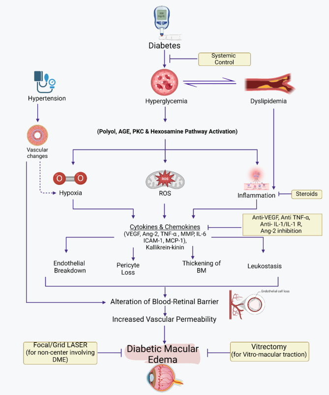
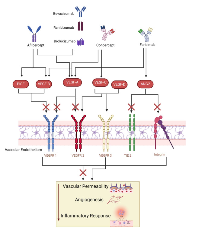
One of the most common health concerns disturbing people within working years globally is diabetes mellitus (DM). One well-known consequence of DM is vascular damage, which can manifest as macro- and microangiopathy affecting the ocular retina. Therefore, Diabetic macular edema (DME) is a major sight-threatening complication of diabetic retinopathy (DR) worldwide. It is the most prevalent cause of significant vision impairment in diabetic patients. Long-term vision loss can be avoided by following early DME treatment guidelines in everyday life. Hence, there are various therapeutic approaches for DME management. Currently, the first-line treatment for DME is anti-VEGF family drugs, such as ranibizumab, brolucizumab, bevacizumab, and aflibercept. Nevertheless, relapses of the disease, inadequate response, and resistance during anti-VEGF therapy are still seen because of the intricate pathophysiological foundation of the disease. Consequently, there is an excellent requirement for therapeutic approaches to advance and become better at controlling diseases more satisfactorily and require fewer treatments overall. We conducted a thorough literature search in the current review to present a comprehensive overview of the primary data about the current DME therapeutic agents. We also covered the novel advances in DME management and probable future treatments being investigated and developed. This review recommended that Large clinical trials should afford sufficient evidence to support these innovative treatment modalities.

Keywords: Anti-VEGFs; Diabetes mellitus; Diabetic macular edema; Proliferative diabetic retinopathy; Selective retinal therapy.


Conflict of interest statement

The authors declare no competing interests.

Figures

Fig. 1
Fig. 1
The pathogenesis of diabetic macular edema and management strategies. Illustrates the progression and effects of DME. It begins with conditions such as Hyperglycemia which could be associated with aggravating factors (i.e. hypertension), leading to Biochemical and Molecular Abnormalities. These abnormalities trigger a series of cellular and molecular reactions, including the generation of Reactive Oxygen Species (ROS) and its effects. These effects include inflammation, endothelial cell junction breakdown, pericyte loss, thickening of the Basement Membrane (BM), and leukostasis. These processes alter the blood-retinal barrier and increase vascular permeability, resulting in Diabetic Macular Edema. The figure indicates current available treatments like Anti-VEGFs, Steroids, Systemic treatments, Focal/Grid Laser (for DME involving the center) and Vitrectomy (for Vitreo-macular traction).
Fig. 2
Fig. 2
The Role of Anti-VEGF Medications in the Management of Diabetic Macular Edema. VEGF is a signal protein that stimulates the formation of blood vessels. When VEGF is overexpressed, it can lead to the vascular permeability and neovascularization seen in DME. Anti-VEGF drugs work by binding to VEGF, inhibiting its ability to bind to its receptors on the surface of endothelial cells. Different anti-VEGF drugs may have different affinities for the various VEGF receptors, which can influence their efficacy in DME management.
Fig. 3
Fig. 3
The summary includes the types and parameters of current laser therapies. (A) When focusing on microaneurysms with focal laser treatment (indicated by a solid orange arrow), it is advisable to apply the laser for a short duration. To plan the laser treatment, images from fluorescein angiography (FA), indocyanine green angiography (ICGA), optical coherence tomography (OCT) B-scan, or OCT angiography (OCTA) would be utilized. (B) Subthreshold laser treatment (indicated by an orange dotted arrow) is aimed at the retinal pigment epithelium (RPE).
Fig. 4
Fig. 4
Optical coherence tomography (OCT) scans were taken of a patient with diabetic macular edema at different time points: (a) before any treatment, (b) 3 months after the first subthreshold micropulse macular laser (SMPL) treatment, (c) 3 months after the second SMPL treatment, (d) an additional 3 months after the second SMPL treatment, and (e) 3 months after the third SMPL treatment.
Fig. 5
Fig. 5
Mechanism of action in laser photobiomodulation.
Fig. 6
Fig. 6
The average difference in visual clarity (with 95% confidence intervals) between the start and 4 months into a preliminary investigation of photobiomodulation (PBM) treatment for diabetic macular edema (Protocol AE).

References

    1. Kim EJ, Lin WV, Rodriguez SM, Chen A, Loya A, Weng CY. Treatment of Diabetic Macular Edema. Curr Diab Rep. Sep. 2019;19(9):68. 10.1007/s11892-019-1188-4. - PubMed
    1. Jain R, Daigavane S. Intravitreal OZURDEX vs. Intravitreal Bevacizumab for Diabetic Macular Edema: a Comprehensive Review. Cureus Mar. 2024. 10.7759/cureus.56796. - PMC - PubMed
    1. Yau JWY, et al. Global prevalence and major risk factors of Diabetic Retinopathy. Diabetes Care. Mar. 2012;35(3):556–64. 10.2337/dc11-1909. - PMC - PubMed
    1. Romero-Aroca P, Baget-Bernaldiz M, Pareja-Rios A, Lopez-Galvez M, Navarro-Gil R, Verges R. Diabetic Macular Edema Pathophysiology: vasogenic versus inflammatory. J Diabetes Res. 2016;2016:1–17. 10.1155/2016/2156273. - PMC - PubMed
    1. Merante D, Menchini F, Truitt KE, Bandello FM. Diabetic Macular Edema. Drug Saf. Aug. 2010;33(8):643–52. 10.2165/11538340-000000000-00000. - PubMed

LinkOut - more resources