. 2024 Oct 28;25(1):387.
doi: 10.1186/s12931-024-03017-4.

Kiss1 receptor knockout exacerbates airway hyperresponsiveness and remodeling in a mouse model of allergic asthma

Affiliations

Kiss1 receptor knockout exacerbates airway hyperresponsiveness and remodeling in a mouse model of allergic asthma

Nilesh Sudhakar Ambhore et al. Respir Res. .

Abstract

Background: In asthma, sex-steroids signaling is recognized as a critical regulator of disease pathophysiology. However, the paradoxical role of sex-steroids, especially estrogen, suggests that an upstream mechanism or even independent of estrogen plays an important role in regulating asthma pathophysiology. In this context, in our previous studies, we explored kisspeptin (Kp) and its receptor Kiss1R's signaling in regulating human airway smooth muscle cell remodeling in vitro and airway hyperresponsiveness (AHR) in vivo in a mouse (wild-type, WT) model of asthma. In this study, we evaluated the effect of endogenous Kp in regulating AHR and remodeling using Kiss1R knockout (Kiss1R-/-) mice.

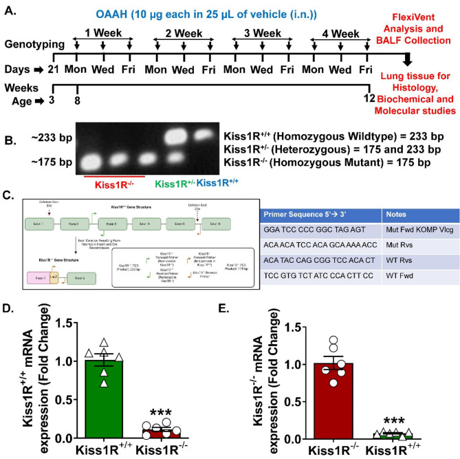
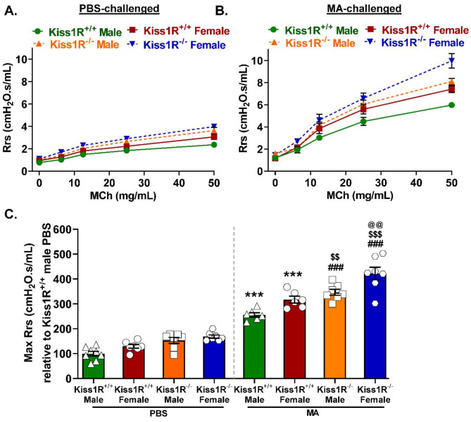
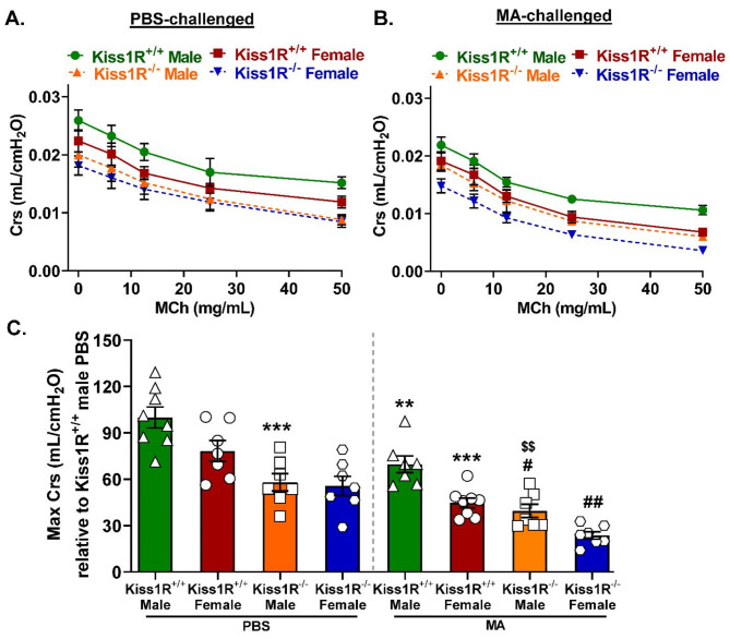
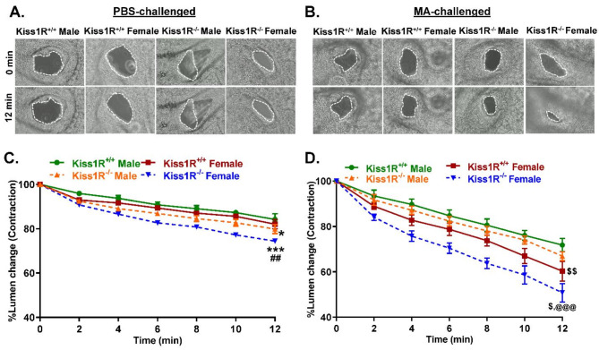
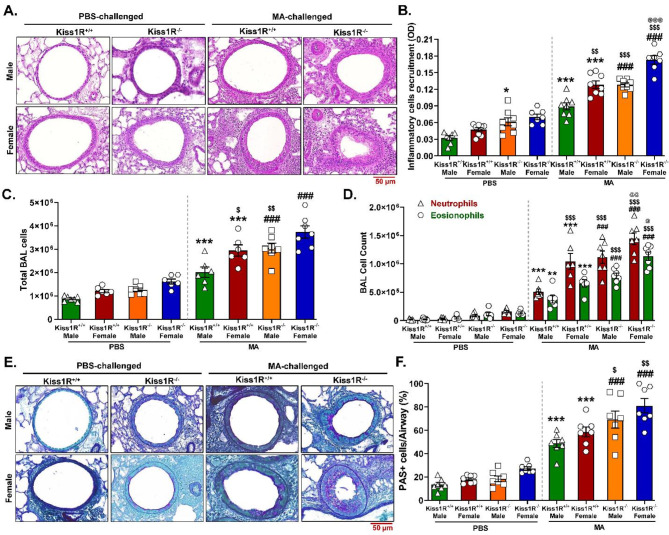
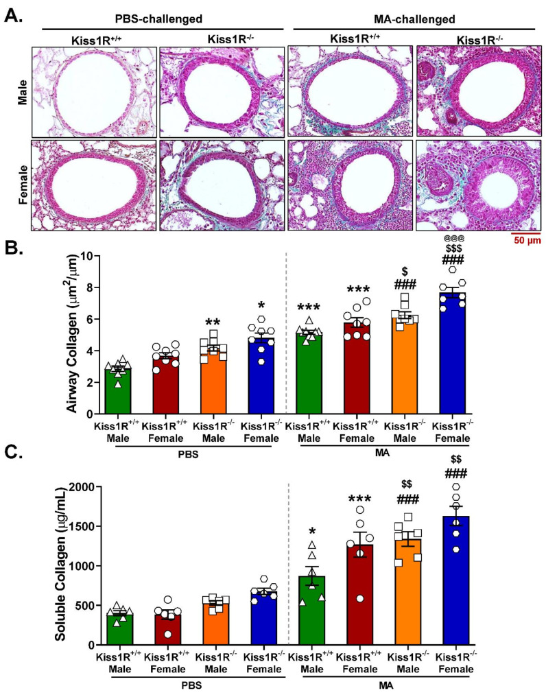
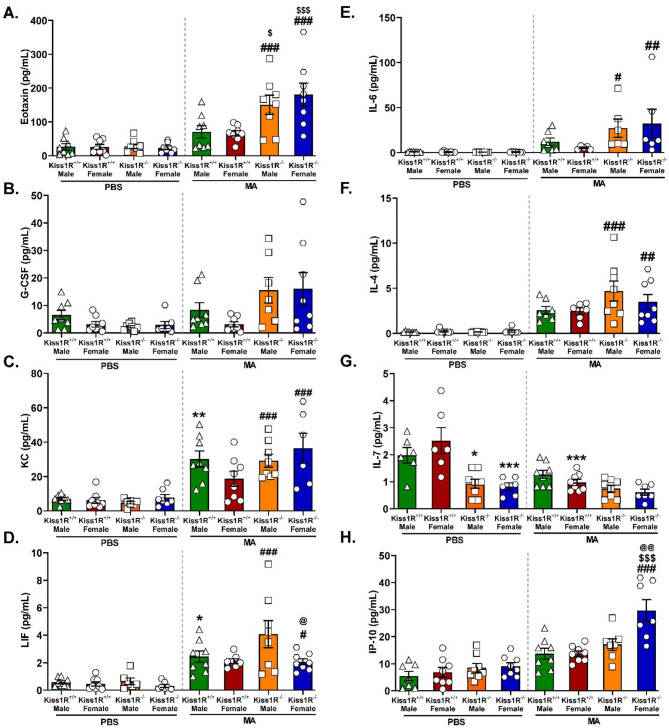
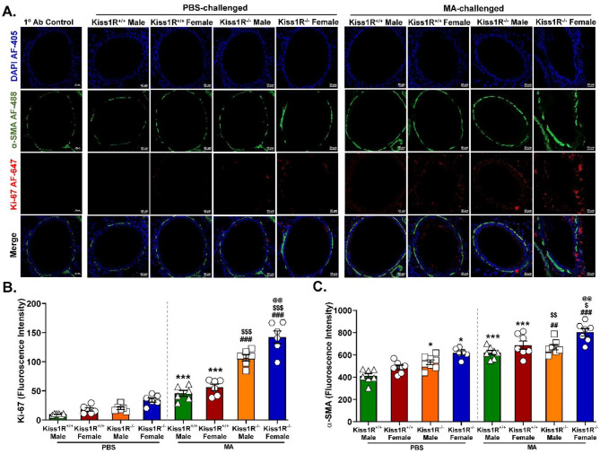
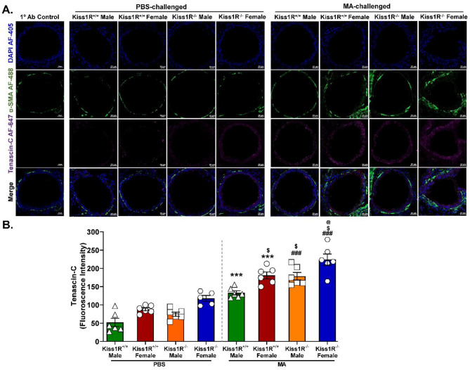
Methods: C57BL/6J WT (Kiss1R+/+) and Kiss1R-/- mice, both male and female, were intranasally challenged with mixed-allergen (MA) and/or phosphate-buffered saline (PBS). We used flexiVent analysis to assess airway resistance (Rrs), elastance (Ers), and compliance (Crs). Following this, broncho-alveolar lavage (BAL) was performed for differential leukocyte count (DLC) and cytokine analysis. Histology staining was performed using hematoxylin and eosin (H&E) for morphological analysis and Masson's Trichrome (MT) for collagen deposition. Additionally, lung sections were processed for immunofluorescence (IF) of Ki-67, α-smooth muscle actin (α-SMA), and tenascin-c.

Results: Interestingly, the loss of Kiss1R exacerbated lung function and airway contractility in mice challenged with MA, with more profound effects in Kiss1R-/- female mice. MA-challenged Kiss1R-/- mice showed a significant increase in immune cell infiltration and proinflammatory cytokine levels. Importantly, the loss of Kiss1R aggravated Th2/Th17 biased cytokines in MA-challenged mice. Furthermore, histology of lung sections from Kiss1R-/- mice showed increased collagen deposition on airway walls and mucin production in airway cells compared to Kiss1R+/+ mice. In addition, immunofluorescence analysis showed loss of Kiss1R significantly aggravated airway remodeling and subsequently AHR.

Conclusions: These findings demonstrate the importance of inherent Kiss1R signaling in regulating airway inflammation, AHR, and remodeling in the pathophysiology of asthma.

Keywords: Airway inflammation; Airway smooth muscle; Asthma; FlexiVent; Lung function; α-smooth muscle actin.


Conflict of interest statement

The authors declare no competing interests.

Figures

Fig. 1
Fig. 1
Schematic of mixed allergen (MA)-induced allergic mouse model of asthma. Genotyping was performed at the age of 3 weeks, and both male and female mice were grouped as Kiss1R+/+ (WT) and Kiss1R−/− (homozygous) according to their respective genotype. Both Kiss1R+/+ and Kiss1R−/− mice challenged with MA regime containing equal amount (10 µg) of each Ovalbumin, Alternaria alternata, Aspergillus fumigatus, and Dermatophagoides pteronyssinus (house dust mite) in 25 µl of Dulbecco’s phosphate-buffered saline (DPBS) on alternative days for 4 weeks (3 days/week). Mice from the control group received only DPBS as a vehicle (A). Representative image of genotyping showing the Kiss1R+/+, Kiss1R+/−, and Kiss1R−/− genotype (B). The illustration in the left panel explains the removal of the Kiss1R gene sequence by inducing a frameshift mutation by Cre recombinase. The primer sequence design specific to Kiss1R+/+ and mutant Kiss1R−/− is provided in the right panel (C). Kiss1R WT mRNA (D) and mutant gene sequence (E) expressions were measured in the lung tissue samples from both Kiss1R+/+ and Kiss1R−/− mice. Data were analyzed using a two-tailed unpaired Student’s t-test. Mean ± SEM (n = 6 mice/group); ***p < 0.001 versus Kiss1R+/+/Kiss1R−/−
Fig. 2
Fig. 2
Loss of Kiss1R aggravates MA-induced total respiratory system resistance (Rrs). Mice from Kiss1R+/+ and Kiss1R−/− groups were exposed to increasing doses (0–50 mg/mL) of nebulized methacholine (MCh) to measure the Rrs after the PBS (A) and MA-challenge (B), using flexiVent analysis. The max Rrs at 50 mg/mL MCh dose was used to compare Kiss1R+/+ vs. Kiss1R−/− and male vs. female mice in both PBS and MA-challenged groups (C). Data were analyzed using one-way ANOVA followed by Tukey’s post hoc test. Mean ± SEM (n = 6–8 mice/group); ***p < 0.001 versus respective Kiss1R+/+ male/female PBS group; ###p < 0.001 versus respective Kiss1R−/− male/female PBS group; $$p < 0.01, $$$p < 0.001 versus respective Kiss1R+/+ male/female MA-challenged group; @@p < 0.01 versus Kiss1R−/− male MA-challenged group
Fig. 3
Fig. 3
Loss of Kiss1R aggravates MA-mediated reduced total respiratory system compliance (Crs). Kiss1R+/+ and Kiss1R−/− mice were exposed to increasing doses (0–50 mg/mL) of nebulized MCh to measure the Crs after the PBS (A) and MA-challenge (B), using flexiVent analysis. The max Crs at 50 mg/mL MCh dose was used to compare Kiss1R+/+ vs. Kiss1R−/− and male vs. female mice in both PBS and MA-challenged groups (C). Data were analyzed using one-way ANOVA followed by Tukey’s post hoc test. Mean ± SEM (n = 7–8 mice/group); **p < 0.01, ***p < 0.001 versus respective Kiss1R+/+ male/female PBS group; #p < 0.05, ##p < 0.01 versus respective Kiss1R−/− male/female PBS group; $$p < 0.01 versus respective Kiss1R+/+ male/female MA-challenged group
Fig. 4
Fig. 4
Kiss1R−/− increases MA-induced airway contractility. Representative images of precision cut lung sections (PCLS) from Kiss1R+/+ and Kiss1R−/− mice challenged with PBS (A) or MA (B). Airway contractility was measured in response to MCh (100 µM) compared to the baseline lumen area. The percent of airway lumen area contraction with MCh was measured for 12 min for each lung section from individual Kiss1R+/+ and Kiss1R−/− mice challenged with PBS (C) or MA (D). Data were analyzed using two-way ANOVA followed by Tukey’s post hoc test. Mean ± SEM (n = 6 mice/group); *p < 0.05, ***p < 0.001 versus respective Kiss1R+/+ male/female PBS group; ##p < 0.01 versus respective Kiss1R−/− male/female PBS group; $p < 0.05, $$p < 0.01 versus respective Kiss1R+/+ male/female MA-challenged group; @@@p < 0.001 versus Kiss1R−/− male MA-challenged group
Fig. 5
Fig. 5
Effect of Kiss1R−/− on MA-induced airway inflammation and goblet cell hyperplasia. Representative image of hematoxylin and eosin (H&E)-stained lung sections from Kiss1R+/+ and Kiss1R−/− mice treated with PBS or MA. Images were captured at 20X magnification, scale bar 50 μm (A). Inflammatory cell recruitments in the respective airways were analyzed by ImageJ and represented as an optical density (OD) value (B). The total number of bronchoalveolar lavage (BAL) cells (C), as well as the number of neutrophils and eosinophils (D) in the BAL fluid of Kiss1R+/+ and Kiss1R−/− mice challenged with PBS or MA were analyzed. Representative image of periodic acid-schiff (PAS)--stained lung sections from Kiss1R+/+ and Kiss1R−/− mice treated with PBS or MA. Images were captured at 20X magnification, scale bar 50 μm (E). The number of PAS-positive cells in the respective airways of Kiss1R+/+ and Kiss1R−/− mice challenged with PBS or MA was measured and normalized to represent a percentage change (F). Data were analyzed using one-way ANOVA followed by Tukey’s post hoc test. Mean ± SEM (n = 6–8 mice/group); *p < 0.05, **p < 0.01, ***p < 0.001 versus respective Kiss1R+/+ male/female PBS group; ###p < 0.001 versus respective Kiss1R−/− male/female PBS group; $p < 0.05, $$p < 0.01, $$$p < 0.001 versus respective Kiss1R+/+ male/female MA-challenged group; @p < 0.05, @@p < 0.01, @@@p < 0.001 versus Kiss1R−/− male MA-challenged group
Fig. 6
Fig. 6
Effect of Kiss1R−/− on MA-induced airway collagen and soluble collagen levels. Representative images of Masson’s trichrome stained lung section from Kiss1R+/+ and Kiss1R−/− mice treated with PBS or MA, scale bar 50 μm (A). The semiquantitative measurement of the area of airway collagen deposition was performed on three fields per lung section, and normalized by an internal perimeter of the respective airway (B). Soluble collagen levels in lung tissue lysates from the Kiss1R+/+ and Kiss1R−/− mice treated with PBS or MA were measured using a Sircol soluble collagen assay kit (C). Data were analyzed using one-way ANOVA followed by Tukey’s post hoc test. Mean ± SEM (n = 6–8 mice/group); *p < 0.05, **p < 0.01, ***p < 0.001 versus respective Kiss1R+/+ male/female PBS group; ###p < 0.001 versus respective Kiss1R−/− male/female PBS group; $p < 0.05, $$p < 0.01, $$$p < 0.001 versus respective Kiss1R+/+ male/female MA-challenged group; @@@p < 0.01 versus Kiss1R−/− male MA-challenged group
Fig. 7
Fig. 7
Effect of Kiss1R−/− on MA-induced airway cytokine and chemokine profiles. The quantification of cytokine and chemokine levels in bronchoalveolar lavage fluids (BALF) collected from the lungs of Kiss1R+/+ and Kiss1R−/− mice treated with MA showed an increase in eotaxin (A), G-CSF (B), KC (C), LIF (D), IL-6 (E), IL-4 (F), IL-7 (G), and IP-10 (H). MA-altered changes in the cytokine and chemokine levels were more profound in Kiss1R−/− male and female mice. Data were analyzed using one-way ANOVA followed by Tukey’s post hoc test. Mean ± SEM (n = 6–8 mice/group); *p < 0.05, **p < 0.01, ***p < 0.001 versus respective Kiss1R+/+ male/female PBS group; #p < 0.05, ##p < 0.01, ###p < 0.001 versus respective Kiss1R−/− male/female PBS group; $p < 0.05, $$$p < 0.001 versus respective Kiss1R+/+ male/female MA-challenged group; @p < 0.05, @@p < 0.01 versus Kiss1R−/− male MA-challenged group
Fig. 8
Fig. 8
Kiss1R−/− increases MA-induced airway cell proliferative markers. Representative immunofluorescence (IF) images of lung sections stained with specific antibodies for Ki-67 (AF-647) and α-SMA (AF-488), with DAPI counterstain (AF-405). Scale bars, 20 μm (A). The quantitative analysis of Ki-67 and α-SMA was performed using Zeiss software. The arithmetic means fluorescence intensity from a minimum of three different regions of the respective airway was used to determine the expression of Ki-67 (B) and α-SMA (C) in lung sections of Kiss1R+/+ and Kiss1R−/− mice treated with PBS or MA. Data were analyzed using one-way ANOVA followed by Tukey’s post hoc test. Mean ± SEM (n = 6–8 mice/group); *p < 0.05, ***p < 0.001 versus respective Kiss1R+/+ male/female PBS group; ##p < 0.01, ###p < 0.001 versus respective Kiss1R−/− male/female PBS group; $p < 0.05, $$p < 0.01, $$$p < 0.001 versus respective Kiss1R+/+ male/female MA-challenged group; @@p < 0.01 versus Kiss1R−/− male MA-challenged group
Fig. 9
Fig. 9
Kiss1R−/− increases MA-induced airway remodeling markers. Representative IF images of lung sections stained with tenascin-c (AF-647) and α-SMA (AF-488) with DAPI counterstain (AF-405). Scale bars, 20 μm (A). The quantitative analysis of tenascin-c was performed using Zeiss software. The arithmetic means fluorescence intensity from a minimum of three different regions of the respective airway was reported to see the changes in tenascin-c (B) expression from the lung sections of Kiss1R+/+ and Kiss1R−/− mice treated with PBS or MA. Data were analyzed using one-way ANOVA followed by Tukey’s post hoc test. Mean ± SEM (n = 5–6 mice/group); ***p < 0.001 versus respective Kiss1R+/+ male/female PBS group; ###p < 0.001 versus respective Kiss1R−/− male/female PBS group; $p < 0.05 versus respective Kiss1R+/+ male/female MA-challenged group; @p < 0.05 versus Kiss1R−/− male MA-challenged group

References

    1. Chiarella SE, Cardet JC, Prakash Y, editors. Sex, cells, and asthma. Mayo Clin Proc. 2021;96(7):1955–1969. - PMC - PubMed
    1. Ambhore NS, Kalidhindi RSR, Sathish V. Sex-Steroid Signaling in Lung diseases and inflammation. Adv Exp Med Biol. 2021;1303:243–73. - PMC - PubMed
    1. Wu M, Wu Q, Liu D, Zu W, Zhang D, Chen L. The global burden of lower respiratory infections attributable to respiratory syncytial virus in 204 countries and territories, 1990–2019: findings from the global burden of Disease Study 2019. Intern Emerg Med. 2024;19(1):59–70. - PubMed
    1. Hernandez-Lara MA, Richard J, Deshpande DA. Diacylglycerol Kinase is a Keystone Regulator of Signaling relevant to the pathophysiology of Asthma. Am J Physiol Lung Cell Mol Physiol. 2024;327(1):L3–18. - PMC - PubMed
    1. Ambhore NS, Balraj P, Pabelick CM, Prakash Y, Sathish V. Estrogen receptors differentially modifies lamellipodial and focal adhesion dynamics in airway smooth muscle cell migration. Mol Cell Endocrinol. 2024;579:112087. - PMC - PubMed

MeSH terms

Substances