. 2024 Oct 24:17:7653-7674.
doi: 10.2147/JIR.S483689. eCollection 2024.

Unleashing AdipoRon's Potential: A Fresh Approach to Tackle Pseudomonas aeruginosa Infections in Bronchiectasis via Sphingosine Metabolism Modulation

Affiliations

Unleashing AdipoRon's Potential: A Fresh Approach to Tackle Pseudomonas aeruginosa Infections in Bronchiectasis via Sphingosine Metabolism Modulation

Jia-Wei Xu et al. J Inflamm Res. .

Abstract

Purpose: Bronchiectasis patients are prone to Pseudomonas aeruginosa infection due to decreased level of sphingosine in airway. Adiponectin receptor agonist AdipoRon activates the intrinsic ceramidase activity of adiponectin receptor 1 (AdipoR1) and positively regulates sphingosine metabolism. This study aimed to investigate the potential therapeutic benefit of AdipoRon against Pseudomonas aeruginosa infection.

Methods: A mouse model of Pseudomonas aeruginosa lung infection and a co-culture model of human bronchial epithelial cells with Pseudomonas aeruginosa were established to explore the protective effect of AdipoRon. Liquid chromatography-mass spectrometry was used to detect the effect of AdipoRon on sphingosine level in lung of Pseudomonas aeruginosa-infected mouse models.

Results: The down-regulation of adiponectin and AdipoR1 in airway of bronchiectasis patients was linked to Pseudomonas aeruginosa infection. By activating AdipoR1, AdipoRon reduced Pseudomonas aeruginosa adherence on bronchial epithelial cells and protected cilia from damage in vitro. With the treatment of AdipoRon, the load of Pseudomonas aeruginosa in lung significantly decreased, and peribronchial inflammatory cell infiltration was lessened in vivo. The reduced level of sphingosine in the airway of Pseudomonas aeruginosa infected mice was replenished by AdipoRon, thus playing a protective role in the airway. Moreover, AdipoRon activated P-AMPKα/PGC1α, inhibited TLR4/P-NF-κB p65, and reduced expression of pro-apoptotic bax. However, the protective effect of AdipoRon on resisting Pseudomonas aeruginosa infection was weakened when AdipoR1 was knocked down.

Conclusion: AdipoRon protects bronchial epithelial cells and lung by enhancing their resistance to Pseudomonas aeruginosa infection. The mechanism might be modulating sphingosine metabolism and activating P-AMPKα/PGC1α while inhibiting TLR4/P-NF-κB p65.

Keywords: AdipoR1; AdipoRon; Pseudomonas aeruginosa; bronchiectasis; sphingosine metabolism.


Conflict of interest statement

The authors declare no conflicts of interest in this work.

Figures

Figure 1
Figure 1
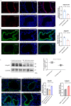
The expression of PPARγ/adiponectin/AdipoR1 was related to Pseudomonas aeruginosa infection in bronchiectasis. (A) The localization of AdipoR1 and adiponectin by immunofluorescence in human primary bronchial epithelial cells differentiated in air-liquid interface. (B) Comparison of ELISA results of adiponectin in BALF between bronchiectasis group and control group (47 patients with bronchiectasis and 22 patients in control group); and adiponectin ELISA results were compared between Pseudomonas aeruginosa-positive bronchiectasis (13 cases) and Pseudomonas aeruginosa-negative bronchiectasis patients (34 cases). (C) The expressions of AdipoR1 and PPARγ in the bronchial epithelium of the patients with bronchiectasis (n = 12) were compared to that of the control group (n = 8) by immunohistochemistry. (D) The expression levels of PPARγ, adiponectin and AdipoR1 in air-liquid differentiated human primary bronchial epithelial cells decreased after infection with Pseudomonas aeruginosa. ***The Mann–Whitney U-test was used to compare the two groups with uneven variance. ***indicates P<0.001.
Figure 2
Figure 2
The expression of adiponectin and AdipoR1 within bronchial epithelium determined in Pseudomonas aeruginosa infected mouse models. (A) Comparison of the expression of adiponectin between Pseudomonas aeruginosa infected mouse models and the uninfected mouse models by immunofluorescence (×200). (B) Comparison of the expression of AdipoR1 between Pseudomonas aeruginosa infected mouse models and the uninfected mouse models by immunofluorescence (×200). (C) Comparison of the expression of AdipoR1 between Pseudomonas aeruginosa infected mouse models and the uninfected mouse models by Western blot. (D) The expression levels of AdipoR1 in the bronchial epithelium of mice in different groups (DMSO control group, AdipoRon high-dose group, AdipoRon low-dose group and adiponectin Acrp30 recombinant protein group) were compared by immunofluorescence (×200). Data are expressed as the means ± standard deviation of three independent experiments. **indicates P<0.01.
Figure 3
Figure 3
AdipoRon reduced bacterial adherence on bronchial epithelial cells and protected cilia from damage. (A and B) Co-culture models of Pseudomonas aeruginosa and air-liquid differentiated human primary bronchial epithelial cells were established. Western blot was used to quantitatively analyze the change in AdipoR1 expression levels with the treatment of AdipoRon. (C and D) Immunofluorescence was used to compare the difference of AdipoR1 expression levels in 5 treatment groups of AdipoRon (×200). (E) Immunofluorescence with an antibody to Pseudomonas aeruginosa (PA1-73116) was used to compare the difference of the adhesion of Pseudomonas aeruginosa on air-liquid differentiated human primary bronchial epithelial cells in 5 treatment groups of AdipoRon (×1000). (F) Scanning electron microscopy was used to observe changes in cilia of cells after the treatment of AdipoRon (red circles indicate interadherent cilia, blue circles indicate broken dissolved cilia, and yellow arrows indicate bacteria). In DMSO control group, bronchial epithelial cells shed a lot of cilia, with more bacteria attached and suspected inflammatory secretions. In 10 μM AdipoRon group, the cilia on the surface of bronchial epithelial cells were broken, shed off, irregular and distributed in clusters, and many bacteria adhered to the cilia. In 50 μM AdipoRon group, the cilia of bronchial epithelial cells were slightly abnormal, and a small number of bacteria were suspected to adhere to the cilia. In 100 μM AdipoRon group, the cilia of the bronchial epithelial cells were slightly abnormal, some of the cilia interweaved into network structure, and no bacterial adhesion was observed. (G) Comparison of the immunofluorescence intensity of the tight junction ZO-1 between the 5 treatment groups of AdipoRon. *indicates P<0.05, **indicates P<0.01.
Figure 4
Figure 4
AdipoRon reduced the load of Pseudomonas aeruginosa in the airway of mice. (A) HE staining was performed on lung tissue specimens from mouse models with different treatment (control group without Pseudomonas aeruginosa infection, DMSO control group with Pseudomonas aeruginosa infection, AdipoRon low-dose (5 mg/kg) pretreatment group with Pseudomonas aeruginosa infection, AdipoRon high-dose (50 mg/kg) pretreatment group with Pseudomonas aeruginosa infection, and ACRP30 recombinant protein pretreatment group with Pseudomonas aeruginosa infection. (B) A semi-quantitative grading method of inflammation score was used to evaluate the severity of peribronchial inflammatory cell infiltration: 0, normal; 1, few cells; 2, a ring of inflammatory cells 1 cell layer deep; 3, a ring of inflammatory cells 2–4 cells deep; 4, a ring of inflammatory cells of > 4 cells deep. The inflammation scores of different groups were compared. (C and D) The CFU of Pseudomonas aeruginosa was counted in the lung tissue homogenates of mice in different treatment groups. (E) The bacterial load of Pseudomonas aeruginosa in the lung tissue of mice in different treatment groups was detected by immunofluorescence with the antibody against Pseudomonas aeruginosa (PA1-73116). Data are expressed as the means ± standard deviation of three independent experiments. **indicates P<0.01.
Figure 5
Figure 5
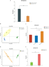
AdipoRon increased sphingosine level in the lung of mouse infection model. (A)The levels of sphingosine were compared between the Pseudomonas aeruginosa-infected mouse model group and the uninfected control group. (B) The metabolites in the lung tissue of mice in 3 groups were compared by PCA analysis: DMSO control group, adipoRon low-dose group and adipoRon high-dose group. (C) The levels of sphingosine in 3 groups were compared. (D) The metabolites in the lung tissue of mice were compared between adiponectin Acrp30 recombinant protein pretreatment group and the DMSO control group by PCA analysis. (E) The levels of sphingosine in 2 groups were compared. ***P<0.001.
Figure 6
Figure 6
AdipoRon activated P-AMPKα/PGC1α, inhibited TLR4/P-NF-κB p65, and reduced expression of bax in Pseudomonas aeruginosa-infected mouse model. (A and B) Western blot was used to quantitatively analyze the expression levels of several major indicators in the lung tissues of mice in three treatment groups (DMSO control group, AdipoRon low-dose group and AdipoRon high-dose group). Image J software was used to analyze the gray levels of different protein imprinting bands and to make statistical analysis. (C and D) Western blot was used to quantitatively analyze the expression levels of several major indexes in the lung tissues of mice in two groups: DMSO control group and Acrp30 recombinant protein pretreatment group. Data are expressed as the means ± standard deviation of three independent experiments. *indicates P<0.05, **indicates P<0.01.
Figure 7
Figure 7
AdipoRon activated P-AMPKα/PGC1α, inhibited TLR4/P-NF-κB p65, and reduced expression of bax in differentiated human primary bronchial epithelial cells. (A) Western blot was used to quantitatively analyze the expression levels of several major indicators of air-liquid differentiated human primary bronchial epithelial cells in 4 treatment groups (DMSO control group, 100 μM AdipoRon group, DMSO control with Pseudomonas aeruginosa infection group, and 100 μM AdipoRon with Pseudomonas aeruginosa infection group). (B and C) Image J Software was used to analyze the gray scale of different Western blot bands and make statistical analysis. (D) Immunofluorescence was used to compare the change in TLR4 and PGC1α in 4 treatment group (×400). (E) Bcl2/bax was quantitatively analyze by Western blot, and the gray scale of the Western blot bands was quantified by Image J software to compare the differences of bcl2, bax and bcl2/bax ratios in 4 groups. (F) The difference of apoptosis rate in 4 groups was compared. (G) TUNEL was used to detect the apoptosis of air-liquid differentiated human bronchial epithelial cells in 4 groups (×200). *indicates P<0.05, **indicates P<0.01.
Figure 8
Figure 8
P-AMPKα/PGC1α was inhibited and TLR4/P-NF-κb p65 was activated after AdipoR1 knockdown by siRNA. (A) Western blot was used to quantitatively analyze the AdipoR1 expression of 16HBE cells in 5 groups (PBS control group, DMSO control group, 10μM AdipoRon group, 50μM AdipoRon group and 100μM AdipoRon group). (B) Comparison of AdipoR1 expression levels by immunofluorescence of 16HBE cells in 5 treatment groups. (CE) qRT-PCR and Western blot were used to analysis the decreased expression of AdipoR1 in 16HBE cells after transfection with AdipoR1 siRNA1 or siRNA2. (C and F) The effect of AdipoRon on the expression of P-AMPKα/PGC1α after AdipoR1 was knocked down was analyzed by Western blot. (G and H) The effect of AdipoRon on the expression level of TLR4/P-NF-κb P65 after AdipoR1 was knocked down was analyzed by Western blot. (I and J) After AdipoR1 was knocked down, the effect of AdipoRon on the expression level of apoptosis marker bcl2/bax was quantitatively analyzed. *indicates P<0.05, **indicates P<0.01.
Figure 9
Figure 9
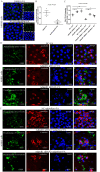
Increased Pseudomonas aeruginosa adhered to 16HBE cells after knockdown of AdipoR1. (A and B) Compared with DMSO control, the addition of 50 μM AdipoRon significantly reduced the amount of Pseudomonas aeruginosa adhered to the surface of 16HBE cells by immunofluorescence (×1000). (C and D) After knockdown of AdipoR1 expression levels by AdipoR1 siRNA1 or AdipoR1 siRNA2, the amount of Pseudomonas aeruginosa adhered to the surface of 16HBE cells was tested by immunofluorescence. (E) After knockdown of AdipoR1 expression levels, the effect of 50μM AdipoRon on the amount of Pseudomonas aeruginosa adhering to the surface of 16HBE cells was analyzed. *indicates P<0.05, **indicates P<0.01.
Figure 10
Figure 10
AdipoRon positively regulates sphingosine metabolic balance by up-regulating AdipoR1 to resisting Pseudomonas aeruginosa infection. The expression of PPARγ in bronchiectasis may be down-regulated by some mechanism, which may result in down-regulation of adiponectin expression and AdipoR1 expression. The decrease of sphingosine increased the susceptibility of airway epithelium to Pseudomonas aeruginosa infection. AdipoRon, an adiponectin receptor agonist, not only positively regulates sphingosine metabolic balance by up-regulating AdipoR1 expression, but also activating the P-AMPK α/PGC1 α pathway, inhibiting TLR4/P-NF-κB p65 pathway, and reducing the expression of pro-apoptosis bax. AdipoRon protects bronchial epithelial cells and lung by enhancing their resistance to Pseudomonas aeruginosa infection.

Similar articles

References

    1. Ebenezer DL, Berdyshev EV, Bronova IA, et al. Pseudomonas aeruginosa stimulates nuclear sphingosine-1-phosphate generation and epigenetic regulation of lung inflammatory injury. Thorax. 2019;74(6):579–591. doi:10.1136/thoraxjnl-2018-212378 - DOI - PMC - PubMed
    1. Gardner AI, Wu Y, Verhaegh R, et al. Interferon regulatory factor 8 regulates expression of acid ceramidase and infection susceptibility in cystic fibrosis. J Biol Chem. 2021;296:100650. doi:10.1016/j.jbc.2021.100650 - DOI - PMC - PubMed
    1. Becker KA, Riethmüller J, Seitz AP, et al. Sphingolipids as targets for inhalation treatment of cystic fibrosis. Adv Drug Deliv Rev. 2018;133:66–75. doi:10.1016/j.addr.2018.04.015 - DOI - PubMed
    1. Becker KA, Verhaegh R, Verhasselt HL, et al. Acid ceramidase rescues cystic fibrosis mice from pulmonary infections. Infect Immun. 2021;89(2):e00677–20. doi:10.1128/IAI.00677-20 - DOI - PMC - PubMed
    1. Gardner AI, Haq IJ, Simpson AJ, et al. Recombinant acid ceramidase reduces inflammation and infection in cystic fibrosis. Am J Respir Crit Care Med. 2020;202(8):1133–1145. doi:10.1164/rccm.202001-0180OC - DOI - PMC - PubMed

LinkOut - more resources