. 2024 Sep 30:15:100400.
doi: 10.1016/j.jpi.2024.100400. eCollection 2024 Dec.

AI drives the assessment of lung cancer microenvironment composition

Affiliations

AI drives the assessment of lung cancer microenvironment composition

Enzo Gallo et al. J Pathol Inform. .

Abstract

Purpose: The abundance and distribution of tumor-infiltrating lymphocytes (TILs) as well as that of other components of the tumor microenvironment is of particular importance for predicting response to immunotherapy in lung cancer (LC). We describe here a pilot study employing artificial intelligence (AI) in the assessment of TILs and other cell populations, intending to reduce the inter- or intra-observer variability that commonly characterizes this evaluation.

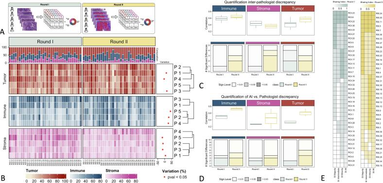
Design: We developed a machine learning-based classifier to detect tumor, immune, and stromal cells on hematoxylin and eosin-stained sections, using the open-source framework QuPath. We evaluated the quantity of the aforementioned three cell populations among 37 LC whole slide images regions of interest, comparing the assessments made by five pathologists, both before and after using graphical predictions made by AI, for a total of 1110 quantitative measurements.

Results: Our findings indicate noteworthy variations in score distribution among pathologists and between individual pathologists and AI. The AI-guided pathologist's evaluations resulted in reduction of significant discrepancies across pathologists: three comparisons showed a loss of significance (p > 0.05), whereas other four showed a reduction in significance (p > 0.01).

Conclusions: We show that employing a machine learning approach in cell population quantification reduces inter- and intra-observer variability, improving reproducibility and facilitating its use in further validation studies.

Keywords: Computer-aided tool; Digital pathology; Lung cancer; Machine learning; NSCLC; Pathology image; QuPath; Tumor-infiltrating lymphocytes; Whole slide images.


Conflict of interest statement

The authors declare the following financial interests/personal relationships which may be considered as potential competing interests: Matteo Pallocca reports a relationship with Dexma srl that includes: consulting or advisory. If there are other authors, they declare that they have no known competing financial interests or personal relationships that could have appeared to influence the work reported in this article.

Figures

Fig. 1
Fig. 1
Graphical Abstract. Depiction of all the steps devoted to the training and the evaluation of the cell classification model. (A) Pre-processing step; (B) training step; (C) test step.
Fig. 2
Fig. 2
(A) Illustration of the pathologist's estimation rounds. Round I and Round II have been performed on the same ROIs with and without the visual support of AI-based cell classification. (B) Pathologist's estimations heatmap. The heatmap shows the percentage of the three cell types assigned by each of the pathologists (P1–P5) for all ROIs. For each ROI, the top annotation describes the average cell composition. Statistically significant percentual variation (across ROIs) in estimates provided by pathologists in Round I and Round II is described and shown in the right annotation. (C) Quantification of inter-pathologist discrepancy in Round I vs Round II. The boxplots represent Kendall correlation coefficients, calculated on the estimates given by all couples of pathologists (across ROIs). The bar plots represent the number of significant Wilcoxon tests performed on the estimates given by all couples of pathologists (across ROIs), at two different p-value thresholds (0.05 and 0.01). (D) Quantification of AI vs. Pathologist discrepancy. The boxplots represent Kendall correlation coefficients, calculated on the estimates given by the AI vs. each of the pathologists (across ROIs). The bar plots represent the number of significant Wilcoxon tests performed on the estimates given by the AI vs. each of the pathologists (across ROIs), at two different p-value thresholds (0.05 and 0.01). (E) Sharing Index (SI) heatmap in Round I and Round II for all three hierarchical positions as well as majority, minority, and intermediate hierarchical position individually. ROIs on the y-axis are sorted from the highest to the lowest global SI. A SI of 1 indicates complete agreement, whereas a SI of 0 indicates the maximum level of discrepancy across pathologists.
Supplementary fig. 1
The illustration shows one of the ROIs used for training and assessment. The sample ROI was used for the sample assignment. The selected nuclei exhibited a Point object within their respective areas. The training ROI (on the right) was employed for performance evaluations and the calculation of classification metrics. In this instance, the Live Prediction was active. The AI classified the nuclei in red as tumoral cells, the nuclei in blue as immune cells, and the nuclei in magenta as stromal cells.

References

    1. Corredor G., Wang X., Zhou Y., et al. Spatial architecture and arrangement of tumor-infiltrating lymphocytes for predicting likelihood of recurrence in early-stage non-small cell lung cancer. Clin Cancer Res Off J Am Assoc Cancer Res. 2019;25(5) doi: 10.1158/1078-0432.CCR-18-2013. - DOI - PMC - PubMed
    1. Geng Y., Shao Y., He W., et al. Prognostic role of tumor-infiltrating lymphocytes in lung cancer: a meta-analysis. Cell Physiol Biochem Int J Exp Cell Physiol Biochem Pharmacol. 2015;37(4) doi: 10.1159/000438523. - DOI - PubMed
    1. Bremnes R., Donnem T., Busund L. Importance of tumor infiltrating lymphocytes in non-small cell lung cancer? Ann Transl Med. 2016;4(7) doi: 10.21037/atm.2016.03.28. - DOI - PMC - PubMed
    1. Bruni D., Angell H., Galon J. The immune contexture and Immunoscore in cancer prognosis and therapeutic efficacy. Nat Rev Cancer. 2020;20(11) doi: 10.1038/s41568-020-0285-7. - DOI - PubMed
    1. Brambilla E., Le Teuff G., Marguet S., et al. Prognostic effect of tumor lymphocytic infiltration in Resectable non-small-cell lung cancer. J Clin Oncol Off J Am Soc Clin Oncol. 2016;34(11) doi: 10.1200/JCO.2015.63.0970. - DOI - PMC - PubMed

LinkOut - more resources