. 2024 Oct 24:19:10725-10743.
doi: 10.2147/IJN.S477159. eCollection 2024.

The Cervical and Meningeal Lymphatic Network as a Pathway for Retrograde Nanoparticle Transport to the Brain

Affiliations

The Cervical and Meningeal Lymphatic Network as a Pathway for Retrograde Nanoparticle Transport to the Brain

Héctor M Ramos-Zaldívar et al. Int J Nanomedicine. .

Abstract

Introduction: The meningeal lymphatic vessels have been described as a pathway that transports cerebrospinal fluid and interstitial fluid in a unidirectional manner towards the deep cervical lymph nodes. However, these vessels exhibit anatomical and molecular characteristics typical of initial lymphatic vessels, with the absence of surrounding smooth muscle and few or absent valves. Given its structure, this network could theoretically allow for bidirectional motion. Nevertheless, it has not been assessed as a potential route for nanoparticles to travel from peripheral tissues to the brain.

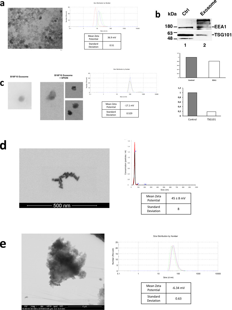
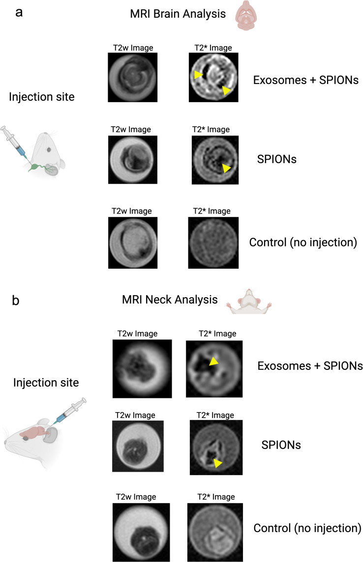
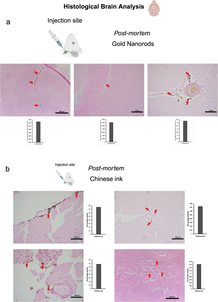
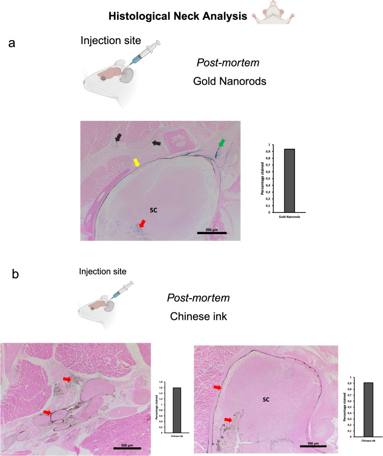
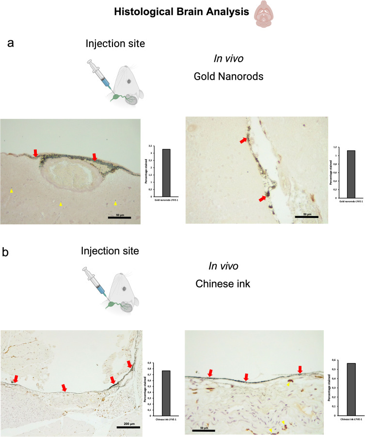
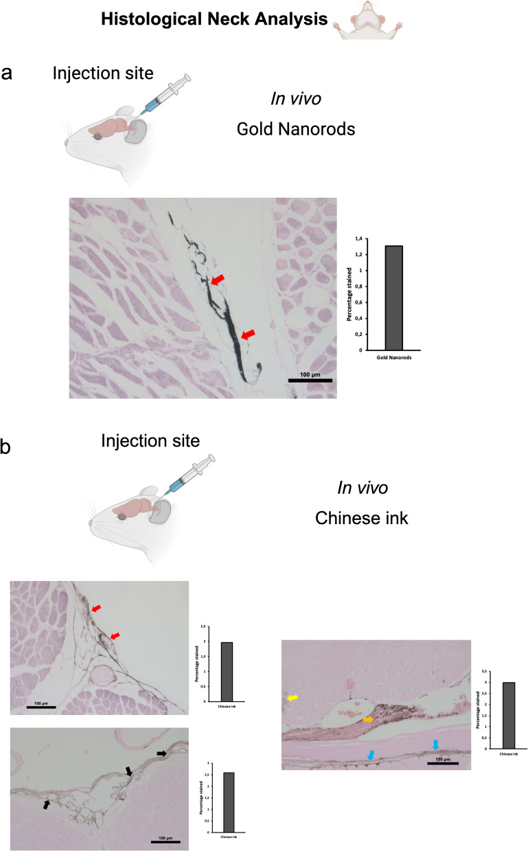
Methods: We employed superparamagnetic iron oxide nanoparticles (SPIONs), exosomes loaded with SPIONs, gold nanorods, and Chinese ink nanoparticles. SPIONs were prepared via chemical coprecipitation, while exosomes were isolated from the B16F10 melanoma cell line through the Exo-Spin column protocol and loaded with SPIONs through electroporation. Gold nanorods were functionalized with polyethylene glycol. We utilized C57BL/6 mice for post-mortem and in vivo procedures. To evaluate the retrograde directional flow, we injected each nanoparticle solution in the deep cervical lymph node. The head and neck were fixed for magnetic resonance imaging and histological analysis.

Results: Here we show that extracellular vesicles derived from the B16F10 melanoma cell line, along with superparamagnetic iron oxide nanoparticles, gold nanorods, and Chinese ink nanoparticles can reach the meningeal lymphatic vessels and the brain of C57BL/6 mice after administration within the deep cervical lymph nodes post-mortem and in vivo, exclusively through lymphatic structures.

Discussion: The functional anatomy of dural lymphatics has been found to be conserved between mice and humans, suggesting that our findings may have significant implications for advancing targeted drug delivery systems using nanoparticles. Understanding the retrograde transport of nanoparticles through the meningeal lymphatic network could lead to novel therapeutic approaches in nanomedicine, offering new insights into fluid dynamics in both physiological and neuropathological contexts. Further research into this pathway may unlock new strategies for treating neurological diseases or enhancing drug delivery to the brain.

Keywords: Chinese ink; SPIONs; extracellular vesicles; gold nanorods; meningeal lymphatic vessels; nanomedicine.


Conflict of interest statement

The authors declare no competing interests in this work.

Figures

Figure 1
Figure 1
Characterization of nanoparticles. (a) STEM visualization of SPIONs; size distribution and zeta potential measured by DLS. (b) Western blot of EV markers EEA1 and TSG101 on exosomes from the B16F10 melanoma cell line. Control was performed using a cellular extract from the B16F10 cell line. Quantification of EEA1 and TSG101 with respect to control is shown. (c) STEM visualization of exosomes with and without SPIONs labeling; size distribution and zeta potential measured by DLS. (d) STEM visualization of gold nanorods; size distribution measured by NTA and zeta potential determined by DLS. (e) STEM visualization of Chinese ink; size distribution and zeta potential measured by DLS.
Figure 2
Figure 2
Directional flow analysis by MRI of SPIONs and SPION-labeled exosomes through the cervical and meningeal lymphatic network. (a) Retrograde Directional Analysis: Brain images reveal the detection of nanoparticles in this region 30 minutes after injection into the deep cervical lymph node (n=3), particularly evident in the T2* map (yellow arrows). These two conditions were compared to control mice with no injected solutions (n=3). (b) Anterograde Directional Analysis: Neck images reveal the detection of nanoparticles in this region 30 minutes after injection into the cisterna magna (n=3), particularly evident in the T2* map (yellow arrows). These two conditions were compared to control mice with no injected solutions (n=3).
Figure 3
Figure 3
Retrograde directional flow analysis by brain histology after post-mortem nanoparticle administration into the deep cervical lymph node. (a) Gold nanorods were identified by the Gold Enhancement technique in the olfactory bulb, the brain parenchyma, and the meningeal lymphatic vessels (red arrows) (n=3). (b) Chinese ink nanoparticles stained the meningeal lymphatic vessels, the brain parenchyma, and the third ventricle wall (red arrows) (n=3).
Figure 4
Figure 4
Anterograde directional flow analysis by brain histology after post-mortem nanoparticle administration into the cisterna magna. (a) Gold Enhancement showed staining of the cervical spinal cord (red arrow), its surrounding subdural space (yellow arrow) and associated peripheral nerves (green arrow). Gold nanoparticles were also detected in cervical lymphatic vessels and connective tissue (black arrows) (n=3). (b) Chinese ink nanoparticles were identified in cervical lymphatic vessels, connective tissue, as well as the cervical spinal cord and its surrounding subdural space (indicated by red arrows) (n=3).
Figure 5
Figure 5
Retrograde directional flow analysis by brain histology after in vivo nanoparticle administration into the deep cervical lymph node. (a) Combined Gold Enhancement and anti-LYVE-1 immunohistochemistry showed gold nanorods within meningeal lymphatic vessels (red arrows) and the brain parenchyma (yellow arrows), with no staining within cerebral arteries (n=3). (b) Meningeal lymphatic vessels stained with anti-LYVE-1 immunohistochemistry and colocalized with Chinese ink nanoparticles (red arrows). Chinese ink was also identified in the brain parenchyma (yellow arrows) (n=4).
Figure 6
Figure 6
Anterograde directional flow analysis by brain histology after in vivo nanoparticle administration into the cisterna magna. (a) Gold Enhancement showed staining of lymphatic vessels in the cervical region (red arrows) (n=3). (b) Chinese ink nanoparticles were identified in the cervical lymphatic vessels (red arrows), the subarachnoid space (black arrows), as well as the cervical spinal cord (yellow arrow), peripheral nerves (Orange arrow), and connective tissue (blue arrows) (n=3).

References

    1. Hershenhouse KS, Shauly O, Gould DJ, Patel KM. Meningeal lymphatics: a review and future directions from a clinical perspective. neuroscience Insights. 2019;14:1179069519889027. doi:10.1177/1179069519889027 - DOI - PMC - PubMed
    1. Jiang H, Wei H, Zhou Y, Xiao X, Zhou C, Ji X. Overview of the meningeal lymphatic vessels in aging and central nervous system disorders. Cell Biosci. 2022;12(1):202. doi:10.1186/s13578-022-00942-z - DOI - PMC - PubMed
    1. Ali Khan A, Mudassir J, Mohtar N, Darwis Y. Advanced drug delivery to the lymphatic system: lipid-based nanoformulations. Int J Nanomed. 2013;8:2733–2744. doi:10.2147/IJN.S41521 - DOI - PMC - PubMed
    1. Louveau A, Smirnov I, Keyes TJ, et al. Structural and functional features of central nervous system lymphatic vessels. Nature. 2015;523(7560):337–341. doi:10.1038/nature14432 - DOI - PMC - PubMed
    1. Aspelund A, Antila S, Proulx ST, et al. A dural lymphatic vascular system that drains brain interstitial fluid and macromolecules. J Exp Med. 2015;212(7):991–999. doi:10.1084/jem.20142290 - DOI - PMC - PubMed

LinkOut - more resources