. 2024 Oct 14:15:1474898.
doi: 10.3389/fphar.2024.1474898. eCollection 2024.

Capsaicin attenuates the effect of inflammatory cytokines in a HaCaT cell model for basal keratinocytes

Affiliations

Capsaicin attenuates the effect of inflammatory cytokines in a HaCaT cell model for basal keratinocytes

Maria Fernanda Cervantes Recalde et al. Front Pharmacol. .

Abstract

Introduction: The resolution of the skin's inflammatory response is only possible if its barrier function is restored. TRPV1 channel activation plays an important role during inflammation but the effect of this activation on the skin barrier under inflammatory conditions has not been clarified. We hypothesize that it could potentially aid the keratinocyte barrier by reducing inflammatory cytokine release and promoting tight junction development.

Methods: To explore the role of TRPV1 activation in inflammation, we designed and optimized an in vitro model of keratinocytes with basal epidermal layer characteristics using HaCaT cells and TNFα to induce inflammation.

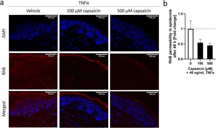
Results: TNFα increased the gene expression of tight junction protein claudin 1 (CLDN1) by at least 2.60 ± 0.16-fold, in a concentration-dependent manner, over a 48 h period. The administration of a capsaicin pre-treatment reduced the CLDN1 expression to 1.51 ± 0.16-fold during the first 6 h after TNFα induction, whereas IL-8 cytokine release was reduced 0.64 ± 0.17-fold. After 48 h, CLDN1 protein levels increased by a factor of 6.57 ± 1.39 compared to cells only treated with TNFα.

Discussion: These results suggest that activation of TRPV1 by capsaicin can potentiate the increase in CLDN1 expression and CLDN1 protein synthesis induced by TNFα in cultured keratinocytes, while reducing the release of IL-8.

Keywords: TRPV1; capsaicin; inflammation; permeability; skin.


Conflict of interest statement

Authors JH and DS were employed by Symrise AG. Authors CG and MM were employed by Symrise Srl. The remaining authors declare that the research was conducted in the absence of any commercial or financial relationships that could be construed as a potential conflict of interest.

Figures

FIGURE 1
FIGURE 1
Characterization of capsaicin pre-treatment of skin biopsies and its effect on TNFα-induced inflammation. Epidermal explants of a healthy adult woman, pre-treated with 100 μM, 500 µM or without capsaicin for 24 h, were incubated with 40 ng/mL TNFα for 48 h before measuring the permeability of a RhB dye through the stratum corneum (SC) using fluorescence microscopy. (A) Images show the permeability of the RhB dye on skin biopsies exposed to vehicle or different concentrations of capsaicin before TNFα-treatment into the inner layers of the epidermis. (B) Average fluorescence readings of RhB dye inside the epidermis of capsaicin pre-treated skin biopsies (black) presented as fold change of the non-pretreated control biopsies (white). (Statistics: mean fold change to non-treated control +SEM; technical replicates: 6, biological replicates: 1).
FIGURE 2
FIGURE 2
Time dependence of the TNFα-induced inflammatory response in HaCaT keratinocytes. (A) TNFα-induced CXCL8 gene expression peaks at early time points (solid line), subsiding with time which contrasts with the steadily increasing IL-8 release (dashed line) that reaches a maximum at the 48 h time point. Control values for CXCL8 and IL-8 release are 1.00 ± 0.06 and 0.11 ± 0.05 pg/mL respectively (B) OCLN expression (dashed line) is significantly increased at early stages of the TNFα response but returns to baseline levels with time. CLDN1 gene expression (solid line) remains steadily upregulated over the whole course of the measurement seeing an increase at 48 h. CLDN2 expression (dotted line) is rather unaffected for the first 24 h to see a significant decrease thereafter. Control values for OCLN, CLDN1 and CLDN2 are 1.00 ± 0.05, 1.00 ± 0.05 and 1.00 ± 0.03, respectively. CXCL8, OCLN, CLDN1 and CLDN2 gene expression were measured using RT-qPCR and IL-8 release was measured using ELISA. (Statistics: mean ± SEM; technical replicates: 3-6, biological replicates: 3-5; Brown-Forsythe and Welch ANOVA with Dunnett’s T3 multiple comparisons post hoc test, (A) ****p < 0.0001 for significance in CXCL8 expression (solid line) and ++++ p < 0.0001 for significance in the IL-8 release (dashed line); (B) ****p < 0.0001 for significance in CLDN1 expression (solid line), ++++ p < 0.0001 for significance in OCLN expression (dashed line), # p < 0.5 for significance in CLDN2 expression (dotted line).
FIGURE 3
FIGURE 3
Increasing concentrations of TNFα promote (A) CXCL8 gene expression and (B) IL-8 release after an incubation period of 48 h. This is accompanied by an equally concentration dependent increase in (C) CLDN1 gene expression. Expression of (D) CLDN2 does not show a concentration dependent increase but is rather stable during the 48 h period with the exception of significant downregulation using 5 ng/mL TNFα. (E) Expression of OCLN was similar to the control over all concentrations. CXCL8, CLDN1, CLDN2 and OCLN gene expression were measured using RT-qPCR and IL-8 release was measured using ELISA. Data is presented as fold change to non-treated control. White bars represent the non-treated control and black bars represent TNFα treatment at different concentrations. (Statistics: mean +SEM; technical replicates: 3-6, biological replicates: 3-5; (A–B) Kruskal Wallis test with Dunn’s multiple comparisons post hoc test **p < 0.01, ***p < 0.001, ****p < 0.0001; (C–E) Brown-Forsythe and Welch ANOVA with Dunnett’s T3 multiple comparisons post hoc test, *p < 0.05, ****p < 0.0001).
FIGURE 4
FIGURE 4
TNFα-induced increase in CLDN1 protein abundance in HaCaT keratinocytes. TNFα increased claudin-1 protein abundance at (A) 6 h and (B) 48 h. Claudin-1 protein abundance was measured with fluorescence microscopy. Data is presented as fold change to non-treated control. White bars represent the non-treated control and black bars represent TNFα treatment at different concentrations. (Statistics: mean +SEM; technical replicates: 3-6, biological replicates: 3-5; Mann-Whitney t-test, ***p < 0.001, ****p < 0.0001).
FIGURE 5
FIGURE 5
Capsaicin regulates the TNFα-induced inflammatory response in HaCaT keratinocytes. (A, B) Cells pretreated with 10 µM capsaicin for 24 h did not show any modulation of the TNFα response with regards to CXCL8 gene expression but (C, D) IL-8 release was significantly attenuated after 6 h and remained lower than in cells that were not pre-treated over 48 h (E, F) CLDN1 gene expression was significantly enhanced in pre-treated cells 6 h after start of the TNFα treatment but the effect subsided with time. (G, H) An increase of CLDN1 protein abundance was evident after 48 h in comparison to the non pre-treated control. CXCL8 and CLDN1 gene expression obtained with real-time PCR and IL-8 release using ELISA. CLDN1 protein was imaged with fluorescence microscopy and fluorescence intensity calculated as fold change of the control. Data presented as the fold change to the non pre-treated TNFα control. (Statistics: mean +SEM; technical replicates: 3, biological replicates: 3; (A–D, F) Welch’s t-test, ****p < 0.0001; (E, G, H) Mann-Whitney t-test, *p < 0.05, ***p < 0.001).
FIGURE 6
FIGURE 6
Decreased IL-8 release and promotion of CLDN1 after capsaicin treatment is mediated by the TRPV1 channel. (A) Cells pre-treated with capsaicin for 24 h reduced the IL-8 release during the first 6 h of TNFα-induced inflammation. Co-incubation with 1 µM capsazepine during the pre-treatment prevented the attenuation of the IL-8 release. (B, C) Similarly, the promotion of CLDN1 and increased abundance of CLDN1 seen after pre-treatment with 10 µM capsaicin at 6 and 48 h, respectively, was no longer present when 1 µM capsazepine was added in the pre-treatment. IL-8 release assessment was done using ELISA, CLDN1 gene expression measured using real-time PCR and CLDN1 protein was measured with fluorescence microscopy. Data is presented as fold change to capsaicin treated control. The dotted black line shows the effect elicited by TNFα without pre-treatment. (Statistics: mean +SEM; technical replicates: 3, biological replicates: 3; (A, C) Mann-Whitney t-test, **p < 0.01; (B) Welch’s t-test, *p < 0.05).
FIGURE 7
FIGURE 7
CLDN1 accumulates in cell boundaries after capsaicin pre-treatment. HaCaT cells that were pre-treated for 24 h with 10 µM capsaicin before 20 ng/mL TNFα treatment showed increased positioning of CLDN1 protein close to the boundaries of the cell when compared to cells treated solely with TNFα (arrows). Already at 6 h, there is definition of the cell boundaries between the keratinocytes. This effect is further enhanced at 48 h, with CLDN1 seemingly redistributing from the inner part of the cell to the boundaries.

References

    1. Amoozadeh Y., Dan Q., Xiao J., Waheed F., Szászi K. (2015). Tumor necrosis factor-α induces a biphasic change in claudin-2 expression in tubular epithelial cells: role in barrier functions. Am. J. Physiology - Cell Physiology 309, C38–C50. 10.1152/ajpcell.00388.2014 - DOI - PMC - PubMed
    1. Archer C. B. (2010). “Functions of the skin,” in Rook's textbook of dermatology. Editors Burns S. B. T., Cox N., Griffiths C. (Oxford, UK: Blackwell Publishing Ltd.).
    1. Bäsler K., Brandner J. M. (2017). Tight junctions in skin inflammation. Pflügers Archiv - Eur. J. Physiology 469, 3–14. 10.1007/s00424-016-1903-9 - DOI - PubMed
    1. Beck L. A., Cork M. J., Amagai M., Benedetto A. D., Kabashima K., Hamilton J. D., et al. (2022). Type 2 inflammation contributes to skin barrier dysfunction in atopic dermatitis. JID Innov. 2 (2022/09), 100131. 10.1016/j.xjidi.2022.100131 - DOI - PMC - PubMed
    1. Bergmann S., Von Buenau B., Vidal-Y-Sy S., Haftek M., Wladykowski E., Houdek P., et al. (2020). Claudin-1 decrease impacts epidermal barrier function in atopic dermatitis lesions dose-dependently. Sci. Rep. 10, 2024. 10.1038/s41598-020-58718-9 - DOI - PMC - PubMed

LinkOut - more resources