. 2024 Oct 14:15:1440592.
doi: 10.3389/fimmu.2024.1440592. eCollection 2024.

Ischemia-induced endogenous Nrf2/HO-1 axis activation modulates microglial polarization and restrains ischemic brain injury

Affiliations

Ischemia-induced endogenous Nrf2/HO-1 axis activation modulates microglial polarization and restrains ischemic brain injury

Ping-Chang Kuo et al. Front Immunol. .

Abstract

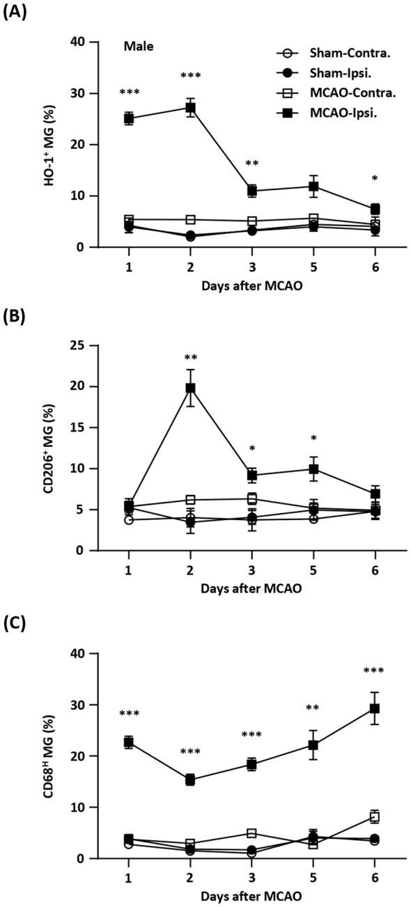
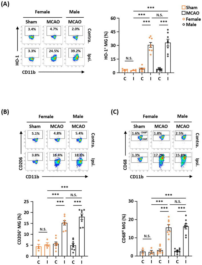
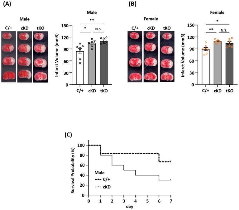
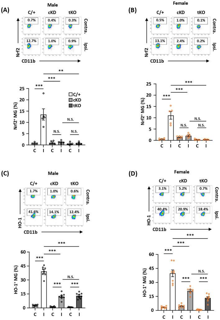
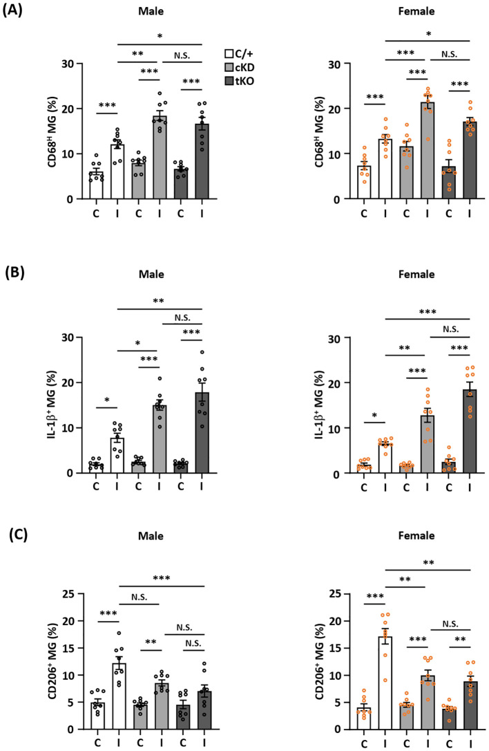
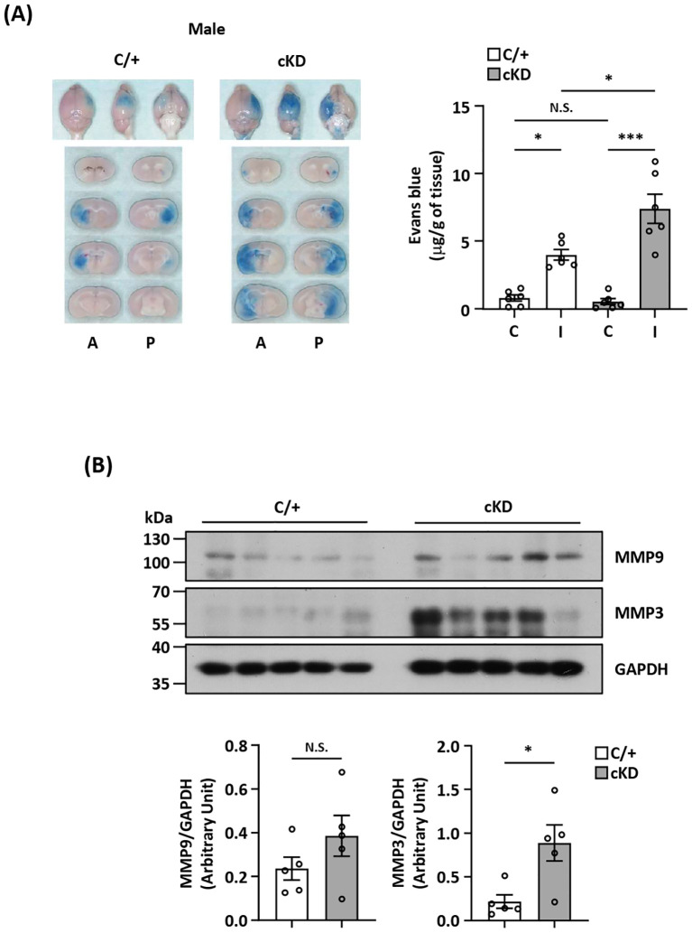
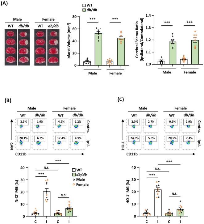
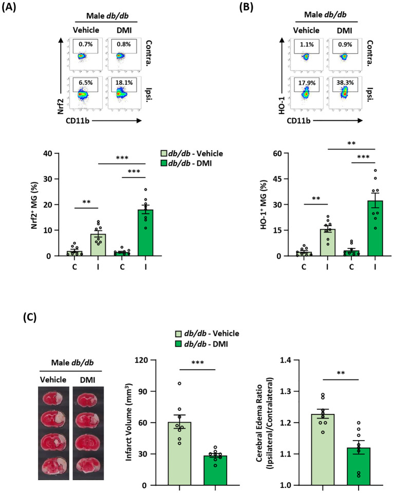
Cerebral ischemic stroke accounts for more than 80% of all stroke cases. During cerebral ischemia, reactive oxygen species produced in the ischemic brain induce oxidative stress and inflammatory responses. Nrf2 is a transcription factor responsible for regulating cellular redox balance through the induction of protective antioxidant and phase II detoxification responses. Although the induction of endogenous Nrf2/HO-1 axis activation has been observed in the ischemic brain, whether ischemia-induced endogenous Nrf2/HO-1 axis activation plays a role in modulating microglia (MG) phenotypes and restraining ischemic brain injury is not characterized and requires further exploration. To investigate that, we generated mice with Nrf2 knockdown specifically in MG to rigorously assess the role of endogenous Nrf2 activation in ischemic brain injury after stroke. Our results showed that MG-specific Nrf2 knockdown exacerbated ischemic brain injury after stroke. We found that Nrf2 knockdown altered MG phenotypes after stroke, in which increased frequency of inflammatory MG and decreased frequency of anti-inflammatory MG were detected in the ischemic brain. Moreover, we identified attenuated Nrf2/HO-1 axis activation led to increased CD68/IL-1β and suppressed CD206 expression in MG, resulting in aggravated inflammatory MG in MG-specific Nrf2 knockdown mice after stroke. Intriguingly, using type II diabetic preclinical models, we revealed that diabetic mice exhibited attenuated Nrf2/HO-1 axis activation in MG and exacerbated ischemic brain injury after stroke that phenocopy mice with MG-specific Nrf2 knockdown. Finally, the induction of exogenous Nrf2/HO-1 axis activation in MG through pharmacological approaches ameliorated ischemic brain injury in diabetic mice. In conclusion, our findings provide cellular and molecular insights demonstrating ischemia-induced endogenous Nrf2/HO-1 axis activation modulates MG phenotypes and restrains ischemic brain injury. These results further strengthen the therapeutic potential of targeting Nrf2/HO-1 axis in MG for the treatment of ischemic stroke and diabetic stroke.

Keywords: Nrf2/HO-1 axis; diabetic stroke; ischemic stroke; microglia; neuroinflammation.


Conflict of interest statement

The authors declare that the research was conducted in the absence of any commercial or financial relationships that could be construed as a potential conflict of interest.

Figures

Figure 1
Figure 1
Temporal expression of HO-1, CD206, and CD68 in MG after ischemic stroke. Male C57BL/6 mice were subjected to sham (n=3/per time point of day 1-6) or 40 min MCAO (n=10/per time point of day 1-5; n=6/day 6). On day 1, 2, 3, 5, and 6 post-injury, mice were sacrificed, and the ischemic brains were harvested. The contralateral (Contra.) and ipsilateral (Ipsi.) hemispheres of sham and MCAO mice were subjected to mononuclear cell isolation. The isolated cells were stained with antibodies against CD45 and CD11b followed by intracellular staining with HO-1, CD206, or CD68 to assess CD45intCD11b+ MG positive for (A) HO-1, (B) CD206, or (C) CD68 expression, respectively. Statistical analysis was comparing the ipsilateral hemisphere of MCAO mice to that of sham controls. *p<0.05; **p<0.01; ***p<0.001 by unpaired t test or Mann-Whitney U test.
Figure 2
Figure 2
Ischemia-induced HO-1, CD206, and CD68 expression in MG is comparable between female and male MCAO mice. Female C57BL/6 mice were subjected to sham (n=4/group) or MCAO (n=8/group). Male C57BL/6 mice were also subjected to MCAO (n=8/group). On day 2 post-injury, the contralateral (Contra.; C) and ipsilateral (Ipsi.; I) hemispheres of sham and MCAO mice were harvested, followed by mononuclear cell isolation. The isolated cells were stained with CD45 and CD11b in the presence of HO-1, CD206, or CD68 to assess CD45intCD11b+ MG positive for (A) HO-1, (B) CD206, or (C) CD68 expression, respectively. The gating of CD68 low (CD68L) was based on the basal level of CD68 expression in MG in sham controls compared to isotype, and the expression level of CD68 higher than CD68L was then determined as CD68 high (CD68H). ***p<0.001; N.S.: no significant difference by two-way ANOVA.
Figure 3
Figure 3
MG-specific Nrf2 knockdown exacerbates ischemic brain injury after stroke. (A) Male and (B) female Cx3cr1CreERT2/+ (C/+), Nrf2fl/fl-Cx3cr1CreERT2/+ (conditional knockdown; cKD), and Nrf2-/- (total knockout; tKO) mice were subjected to MCAO (n=7/group). On day 2 post-injury, the ischemic brains were harvested and subjected to TTC staining. One representative TTC-stained brain sample from each group is shown, and the infarct volumes were measured. *p<0.05; **p<0.01; N.S.: no significant difference by one-way ANOVA. (C) Male C/+ (n=6) and cKD (n=10) mice were subjected to MCAO and monitored to assess the survival rate from day 0 to day 7 post-injury.
Figure 4
Figure 4
MG-specific Nrf2 knockdown results in attenuated HO-1 expression in the ischemic brain after stroke. Male and female Cx3cr1CreERT2/+ (C/+), Nrf2fl/fl-Cx3cr1CreERT2/+ (cKD), and Nrf2-/- (tKO) mice were subjected to MCAO. (A, B) At 5 h post-reperfusion, mice were sacrificed, and the contralateral (Contra.; C) and ipsilateral (Ipsi.; I) hemispheres of male and female MCAO mice were harvested, followed by mononuclear cell isolation. The isolated cells were stained with antibodies against CD45 and CD11b followed by intranuclear staining with Nrf2 antibody. The cells were then subjected to flow cytometry analysis to assess CD45intCD11b+ MG positive for Nrf2 expression (n=5/group). (C, D) On day 1 post-injury, the mononuclear cells isolated from the contralateral and ipsilateral hemispheres of male and female MCAO mice were stained with CD45 and CD11b antibodies followed by intracellular staining with HO-1 antibody. The cells were then subjected to HO-1 expression assessment in CD45intCD11b+ MG (n=8/group). **p<0.01; ***p<0.001; N.S.: no significant difference by two-way ANOVA.
Figure 5
Figure 5
MG-specific Nrf2 knockdown modulates inflammatory and anti-inflammatory phenotypes of MG after ischemic stroke. Male and female Cx3cr1CreERT2/+ (C/+), Nrf2fl/fl-Cx3cr1CreERT2/+ (cKD), and Nrf2-/- (tKO) mice were subjected to MCAO. (A) On day 1 post-injury, the contralateral (C) and ipsilateral (I) hemispheres of MCAO mice were harvested, followed by mononuclear cell isolation. The isolated cells were stained with CD45 and CD11b antibodies followed by intracellular staining with CD68 antibody to determine CD45intCD11b+ MG positive for CD68 expression (n=8/group). (B) The isolated cells were ex vivo cultured in the presence of Golgi Plug for 4.5 h followed by surface staining with CD45 and CD11b antibodies. Following fixation and permeabilization, cells were stained with IL-1β antibody to assess the intracellular IL-1β expression in CD45intCD11b+ MG by flow cytometry (n=8/group). (C) On day 2 post-injury, the isolated cells were stained with CD45 and CD11b antibodies followed by intracellular staining with CD206 antibody to assess CD206-expressing CD45intCD11b+ MG (n=8/group). The representative flow cytometry figures are shown in Supplementary Figure 2 . *p<0.05; **p<0.01; ***p<0.001; N.S.: no significant difference by two-way ANOVA.
Figure 6
Figure 6
MG-specific Nrf2 knockdown aggravates BBB disruption after ischemic stroke. (A) Cx3cr1CreERT2/+ (C/+) and Nrf2fl/fl-Cx3cr1CreERT2/+ (cKD) mice were subjected to 3 h MCAO followed by 3.5 h reperfusion. One hour prior to sacrifice, mice were i.v. injected Evans blue. At 6.5 h post-injury, the ischemic brains were harvested and subjected to sectioning and imaging, and the Evans blue leakage in the contralateral and ipsilateral hemispheres was quantified (n=6/group). Representative ischemic brain images of C/+ and cKD MCAO mice displaying Evans blue extravasation are shown, and the amount of Evans blue extravasation of the contralateral (C) and ipsilateral (I) hemispheres was also quantified. A, anterior surface; P, posterior surface. *p<0.05; ***p<0.001; N.S.: no significant difference by two-way ANOVA. (B) The ipsilateral hemispheres harvested from C/+ and cKD MCAO mice were subjected to western blot analysis for MMP9 and MMP3 expression. The levels of MMP9 and MMP3 expression were also quantified (n=5/group). *p<0.05; N.S.: no significant difference by unpaired t test.
Figure 7
Figure 7
Diabetic stroke exhibits attenuated Nrf2/HO-1 expression in MG and exacerbated ischemic brain injury. (A) Male and female WT and Leprdb/db (db/db) mice were subjected to MCAO and sacrificed at 5 h post-reperfusion. The ischemic brains were harvested and subjected to TTC staining. One representative TTC-stained brain sample from each group is shown, and the infarct volumes were measured. The edema ratio was also calculated (n=8/group). ***p<0.001 by unpaired t test. (B, C) The ischemic brains harvested from male and female WT and db/db MCAO mice were subjected to mononuclear cell isolation. The isolated cells were then stained with CD45 and CD11b antibodies followed by intranuclear staining or intracellular staining with Nrf2 or HO-1 antibodies to determine CD45intCD11b+ MG positive for (B) Nrf2 or (C) HO-1 expression, respectively (n=5/sex/group). ***p<0.001; N.S.: no significant difference by two-way ANOVA.
Figure 8
Figure 8
Pharmacological activation of exogenous Nrf2/HO-1 axis in MG ameliorates diabetes-exacerbated ischemic brain injury. Male Leprdb/db (db/db) mice were subjected to MCAO and then treated with vehicle (n=8) or DMI (n=8) at 1 h post-reperfusion. At 15 h post-injury, the ischemic brains were harvested, followed by mononuclear cell isolation. (A, B) The isolated cells were then stained with antibodies against CD45 and CD11b in the presence of Nrf2 or HO-1 antibodies to assess CD45intCD11b+ MG positive for (A) Nrf2 or (B) HO-1 expression, respectively. **p<0.01; ***p<0.001 by two-way ANOVA. (C) The ischemic brains were harvested from vehicle- and DMI-treated db/db MCAO mice at 15 h post-injury and subjected to TTC staining (n=8/group). One representative TTC-stained brain sample from vehicle- and DMI-treated db/db MCAO mice is shown. The infarct volumes were measured, and the edema ratio was also calculated (n=8/group). **p<0.01; ***p<0.001 by unpaired t test.

References

    1. Lakhan SE, Kirchgessner A, Hofer M. Inflammatory mechanisms in ischemic stroke: therapeutic approaches. J Transl Med. (2009) 7:97. doi: 10.1186/1479-5876-7-97 - DOI - PMC - PubMed
    1. Saini V, Guada L, Yavagal DR. Global epidemiology of stroke and access to acute ischemic stroke interventions . Neurology. (2021) 97:S6–S16. doi: 10.1212/WNL.0000000000012781 - DOI - PubMed
    1. Tsivgoulis G, Katsanos AH, Sandset EC, Turc G, Nguyen TN. Thrombolysis for acute ischaemic stroke: current status and future perspectives. Lancet Neurol. (2023) 22:418–29. doi: 10.1016/S1474-4422(22)00519-1 - DOI - PubMed
    1. Orellana-Urzua S, Rojas I, Libano L, Rodrigo R. Pathophysiology of ischemic stroke: role of oxidative stress. Curr Pharm Des. (2020) 26:4246–60. doi: 10.2174/1381612826666200708133912 - DOI - PubMed
    1. Shah IM, Macrae IM, Di Napoli M. Neuroinflammation and neuroprotective strategies in acute ischaemic stroke - from bench to bedside. Curr Mol Med. (2009) 9:336–54. doi: 10.2174/156652409787847236 - DOI - PubMed

LinkOut - more resources