. 2024 Nov 6;15(21):4075-4081.
doi: 10.1021/acschemneuro.4c00501. Epub 2024 Oct 29.

Cholesterol Accelerates Aggregation of α-Synuclein Simultaneously Increasing the Toxicity of Amyloid Fibrils

Affiliations

Cholesterol Accelerates Aggregation of α-Synuclein Simultaneously Increasing the Toxicity of Amyloid Fibrils

Mikhail Matveyenka et al. ACS Chem Neurosci. .

Abstract

A hallmark of Parkinson disease (PD) is a progressive degeneration of neurons in the substantia nigra pars compacta, hypothalamus, and thalamus. Although the exact etiology of irreversible neuronal degeneration is unclear, a growing body of experimental evidence indicates that PD could be triggered by the abrupt aggregation of α-synuclein (α-Syn), a small membrane protein that is responsible for cell vesicle trafficking. Phospholipids uniquely alter the rate of α-Syn aggregation and, consequently, change the cytotoxicity of α-Syn oligomers and fibrils. However, the role of cholesterol in the aggregation of α-Syn remains unclear. In this study, we used Caenorhabditis elegans that overexpressed α-Syn to investigate the effect of low (15%), normal (30%), and high (60%) concentrations of cholesterol on α-Syn aggregation. We found that an increase in the concentration of cholesterol in diets substantially shortened the lifespan of C. elegans. Using biophysical methods, we also investigated the extent to which large unilamellar vesicles (LUVs) with low, normal, and high concentrations of cholesterol altered the rate of α-Syn aggregation. We found that only lipid membranes with a 60% concentration of cholesterol substantially accelerated the rate of protein aggregation. Cell assays revealed that α-Syn fibrils formed in the presence of LUVs with different concentrations of cholesterol exerted very similar levels of cytotoxicity to rat dopaminergic neurons. These results suggest that changes in the concentration of cholesterol in the plasma membrane, which in turn could be caused by nutritional preferences, could accelerate the onset and progression of PD.

Keywords: Caenorhabditis elegans; ROS; cholesterol; fibrils; α-synuclein.


Conflict of interest statement

The authors declare no competing financial interest.

Figures
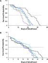
Figure 1
Figure 1
Lipids alter the toxicity of α-Syn aggregates in C. elegans. Kaplan–Meier survival probability curves for NL5901 (A) and N2 WT (B) C. elegans with dietary lipid supplementation of LUVs with 60:40 Cho/DPPC (purple), 30:70 Cho/DPPC (blue), 15:85 Cho/DPPC (light blue), and DPPC (green), as well as lipid-free environment (black). Dashed line (---) indicates p50 of the survival probability.
Figure 2
Figure 2
Lipids alter the concentration of α-Syn in NL5901 C. elegans. Histograms of ELISA of α-Syn possessed by NL5901 C. elegans that were kept on lipid-free diet (black), as well as diet supplied with 60:40 Cho/DPPC (purple), 30:70 Cho/DPPC (blue), 15:85 Cho/DPPC (light blue), and DPPC (green). According to a one-way ANOVA, *P < 0.05, **P < 0.01, ***P < 0.001; NS is nonsignificant difference.
Figure 3
Figure 3
Lipids alter rates of α-Syn aggregation. ThT kinetics (A) with corresponding tlag (B) and t1/2 (C) of α-Syn aggregation in the lipid-free environment (red), as well as in the presence of LUVs that contained 60:40 (purple), 30:70 (blue), 15:85 (light blue) of Cho/DPPC, and 100% DPPC (green). Lag-phase time (tlag) corresponds to 10% increase in the ThT intensity, whereas half-time (t1/2) corresponds to 50% of the maximal ThT intensity observed in the kinetic measurements. Excitation 450 nm; emission 488 nm. According to a one-way ANOVA, **P < 0.01, NS is nonsignificant difference.
Figure 4
Figure 4
Morphological examination and structural characterization of α-Syn aggregates grown in the presence of lipids and in the lipid-free environment. AFM images (A) of α-Syn aggregates formed in the lipid-free environment, as well as in the presence of LUVs that contained 60:40, 30:70, 15:85 of Cho/DPPC, and 100% DPPC. Scale bars are 500 nm. Histogram (B) of height distribution of protein aggregates observed in α-Syn (red), α-Syn:(Cho/DPPC (15:85)) (light blue), α-Syn:(Cho/DPPC (30:70)) (blue), α-Syn:(Cho/DPPC (60:40)) (purple), and α-Syn:DPPC (green). Same number of protein aggregates was analyzed in each sample. FTIR (C) spectra of α-Syn aggregates grown in the lipid-free environment (red), as well as in the presence of LUVs that contained 60:40 (purple), 30:70 (blue), 15:85 (light blue) of Cho/DPPC, and 100% DPPC (green).
Figure 5
Figure 5
Histograms of LDH (A), ROS (B), and JC-1 (C) toxicity assays of α-Syn aggregation in the lipid-free environment (red), as well as in the presence of LUVs that contained 60:40, 30:70, and 15:85 of Cho/DPPC, as well as 100% DPPC (green). Black asterisks (*) show a significant level of differences between α-Syn and α-Syn aggregates grown in the presence of lipids, as well as between lipid samples and the control. Red asterisks (*) show a significant level of differences between α-Syn aggregates grown in the presence of and absence of lipids. According to a one-way ANOVA, *P < 0.05, **P < 0.01, ***P < 0.001, NS is nonsignificant difference.

References

    1. Chen J. Parkinson’s disease: health-related quality of life, economic cost, and implications of early treatment. Am. J. Manag. Care 2010, 16, S87–S93. - PubMed
    1. Bengoa-Vergniory N.; Roberts R. F.; Wade-Martins R.; Alegre-Abarrategui J. Alpha-synuclein oligomers: a new hope. Acta Neuropathol. 2017, 134 (6), 819–838. 10.1007/s00401-017-1755-1. - DOI - PMC - PubMed
    1. Shahmoradian S. H.; Lewis A. J.; Genoud C.; Hench J.; Moors T. E.; Navarro P. P.; Castano-Diez D.; Schweighauser G.; Graff-Meyer A.; Goldie K. N.; et al. Lewy pathology in Parkinson’s disease consists of crowded organelles and lipid membranes. Nat. Neurosci. 2019, 22 (7), 1099–1109. 10.1038/s41593-019-0423-2. - DOI - PubMed
    1. Spillantini M. G.; Schmidt M. L.; Lee V. M.; Trojanowski J. Q.; Jakes R.; Goedert M. α-Synuclein in Lewy bodies. Nature 1997, 388 (6645), 839–840. 10.1038/42166. - DOI - PubMed
    1. Baba M.; Nakajo S.; Tu P. H.; Tomita T.; Nakaya K.; Lee V. M.; Trojanowski J. Q.; Iwatsubo T. Aggregation of alpha-synuclein in Lewy bodies of sporadic Parkinson’s disease and dementia with Lewy bodies. Am. J. Pathol. 1998, 152 (4), 879–884. - PMC - PubMed

Publication types

MeSH terms