. 2024 Dec 15;13(12):bio060380.
doi: 10.1242/bio.060380. Epub 2024 Nov 26.

Disrupting the interaction between AMBRA1 and DLC1 prevents apoptosis while enhancing autophagy and mitophagy

Affiliations

Disrupting the interaction between AMBRA1 and DLC1 prevents apoptosis while enhancing autophagy and mitophagy

Kate Hawkins et al. Biol Open. .

Abstract

AMBRA1 has critical roles in autophagy, mitophagy, cell cycle regulation, neurogenesis and apoptosis. Dysregulation of these processes are hallmarks of various neurodegenerative diseases and therefore AMBRA1 represents a potential therapeutic target. The flexibility of its intrinsically disordered regions allows AMBRA1 to undergo conformational changes and thus to perform its function as an adaptor protein for various different complexes. Understanding the relevance of these multiple protein-protein interactions will allow us to gain information about which to target pharmacologically. To compare potential AMBRA1 activation strategies, we have designed and validated several previously described mutant constructs in addition to characterising their effects on proliferation, apoptosis, autophagy and mitophagy in SHSY5Y cells. AMBRA1TAT, which is a mutant form of AMBRA1 that cannot interact with DLC1 at the microtubules, produced the most promising results. Overexpression of this mutant protected cells against apoptosis and induced autophagy/mitophagy in SHSY5Y cells in addition to enhancing the switch from quiescence to proliferation in mouse neural stem cells. Future studies should focus on designing compounds that inhibit the protein-protein interaction between AMBRA1/DLC1 and thus have potential to be used as a drug strategy for neurodegeneration.

Keywords: Apoptosis; Autophagy; Mitophagy; Neurodegeneration.


Conflict of interest statement

Competing interests All authors are employees and shareholders of Merck & Co., Inc., Rahway, NJ, USA.

Figures
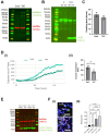
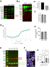
Fig. 1.
Fig. 1.
AMBRA1TAT SHSY5Y validation and characterisation. (A) Western blot of AMBRA1TAT and EmV (control) SHSY5Y cells to show AMBRA1 expression. (B) Western blot of DLC1 protein expression in Co-IP samples of AMBRA1TAT and AMBRA1WT SHSY5Y cells. (C) AMBRA1TAT and EmV SHSY5Y doubling time measured in the Incucyte over a period of multiple days. (D) (i) AMBRA1TAT and EmV SHSY5Ys stained with Caspase-3/7 Green and NucRed and imaged in the Incucyte for 130 h in growth media. Caspase levels were normalised to the phase area of each image. (ii) Sample mean±s.d. (E) Western blot analysis of LC3-II expression in AMBRA1TAT and EmV SHSY5Y cells treated with bafilomycin in growth media for 4 h. (F) Normalized quantification of the mitophagy index of AMBRA1TAT and EmV SHSY5Y cells transduced with mtKeima lentivirus and treated with 1uM CCCP. Scale bar: 50 µm. Sample mean±s.d.***P<0.0005, ****P<0.0001 (two-tailed unpaired t-test). All data presented is representative of three experimental repeats. N=3.
Fig. 2.
Fig. 2.
AMBRA1WD40 SHSY5Y validation and characterisation. (A) Western blot of AMBRA1WD40 and EmV SHSY5Y cells to show AMBRA1 expression. (B) (i-ii) Western blot of DDB1 protein in Co-IP samples of AMBRA1WD40 and AMBRA1WT SHSY5Y cells. (C) AMBRA1WD40 and EmV SHSY5Y cells doubling time measured in the Incucyte over a period of multiple days. (D) (i) AMBRA1WD40 and EmV SHSY5Y cells stained with Caspase-3/7 Green and NucRed and imaged in the Incucyte for approximately 50 h in growth media. Caspase levels were normalised to the phase area of each image. (ii) Sample mean±s.d. (E) Western blot analysis of LC3-II expression in AMBRA1WD40 and EmV SHSY5Y cells treated with bafilomycin in growth media for 4 h. (F) Normalized quantification of the mitophagy index of AMBRA1WD40 and EmV SH-SY5Y cells transduced with mtKeima virus and treated with 1uM CCCP. Sample mean±s.d. *P<0.05, **P<0.01, scale bar: 50 µm. All data presented is representative of three experimental repeats. N=3.
Fig. 3.
Fig. 3.
AMBRA1S1014 SHSY5Y validation and characterisation. (A) Western blot of AMBRA1S1014 and EmV SHSY5Y cells to show AMBRA1 expression. Note that this is from the same blot as Fig. 2A. (B) Western blot of LC3 protein in Co-IP samples of AMBRA1S1014 and AMBRA1WT SHSY5Y cells. (C) AMBRA1S1014 and EmV SHSY5Y cells doubling time measured in the Incucyte over a period of multiple days. (D) (i) AMBRA1S1014 and EmV SHSY5Y cells stained with Caspase-3/7 Green and NucRed and imaged in the Incucyte for approximately 70 h in growth media. Caspase levels were normalised to the phase area of each image. (ii) Sample mean±s.d. (E) Western blot analysis of LC3-II expression in AMBRA1S1014 and EmV SHSY5Y cells treated with growth media and bafilomycin for 4 h. (F) Normalized quantification of the mitophagy index of AMBRA1S1014 and EmV SH-SY5Y cells transduced with mtKeima virus. Sample mean±s.d.; **P<0.01, ***P<0.0005. All data presented are representative of three experimental repeats. N=3.
Fig. 4.
Fig. 4.
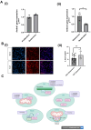
AMBRA1TAT overexpression in mouse neural stem cells. (A) Quantification of the fold change of Ambra1 transcript expression in mNSCs that have been (i) induced to become quiescent and (ii) reactivated from quiescence to proliferation. Two-tailed unpaired t-test where *P<0.05. (Bi-ii) Immunostaining of Hoescht (blue) and Ki-67 (red) in proliferating and quiescent mNSCs. Quiescence was induced using BMP-4. Scale bar: 50 µm. Images taken at 40× magnification. Averages taken from eight fields of view. Error bars±s.e.m. Two-tailed unpaired t-tests where **P<0.01. N=3. (C) Summary of findings. Created with Biorender.com. All data presented is representative of three experimental repeats. N=3.

References

    1. Aman, Y., Schmauck-Medina, T., Hansen, M., Morimoto, R. I., Simon, A. K., Bjedov, I., Palikaras, K., Simonsen, A., Johansen, T., Tavernarakis, N.et al. (2021). Autophagy in healthy aging and disease. Nat. Aging 1, 634-650. 10.1038/s43587-021-00098-4 - DOI - PMC - PubMed
    1. Antonioli, M., Albiero, F., Nazio, F., Vescovo, T., Perdomo, A. B., Corazzari, M., Marsella, C., Piselli, P., Gretzmeier, C., Dengjel, J.et al. (2014). AMBRA1 interplay with cullin E3 ubiquitin ligases regulates autophagy dynamics. Dev. Cell 31, 734-746. 10.1016/j.devcel.2014.11.013 - DOI - PubMed
    1. Chaikovsky, A. C., Li, C., Jeng, E. E., Loebell, S., Lee, M. C., Murray, C. W., Cheng, R., Deumeter, J., Swaney, D. L., Chen, S. H.et al. (2021). The AMBRA1 E3 ligase agaptor regulates the stability of cyclin D. Nature 592, 794-798. 10.1038/s41586-021-03474-7 - DOI - PMC - PubMed
    1. Cianfanelli, V., Fuoco, C., Lorente, M., Salazar, M., Quongamatteo, F., Gherardini, P. F., De Zio, D., Nazio, F., Antionioli, M., D‘Orazio, N.et al. (2015). AMBRA1 links autophagy to cell proliferation and tumorigenesis by promoting c-Myc dephosphorylation and degradation. Nat. Cell Biol. 17, 20-30. 10.1038/ncb3072 - DOI - PMC - PubMed
    1. Di Bartolomeo, S., Corazzari, M., Nazio, F., Oliverio, S., Lisi, G., Antionoli, M., Pagliarini, V., Matteoni, S., Fuoco, C., Giunta, L.et al. (2010). The dynamic interaction of AMBRA1 with the dynein motor complex regulates mammalian autophagy. J. Cell Biol. 191, 155-168. 10.1083/jcb.201002100 - DOI - PMC - PubMed

Publication types

Substances