Comparative Study
. 2024 Nov 5;13(21):e037258.
doi: 10.1161/JAHA.124.037258. Epub 2024 Oct 29.

Characterizing Nonculprit Lesions and Perivascular Adipose Tissue of Patients Following Acute Myocardial Infarction Using Coronary Computed Tomography Angiography: A Comparative Study

Affiliations
Comparative Study

Characterizing Nonculprit Lesions and Perivascular Adipose Tissue of Patients Following Acute Myocardial Infarction Using Coronary Computed Tomography Angiography: A Comparative Study

Xiaomeng Wang et al. J Am Heart Assoc. .

Abstract

Background: The comparison of coronary computed tomography angiography plaques and perivascular adipose tissue (PVAT) between patients with acute myocardial infarction (AMI) posttreatment and patients with stable coronary artery disease is poorly understood. Our objective was to evaluate the differences in coronary computed tomography angiography-quantified plaque and PVAT characteristics in patients post-AMI and identify signs of residual inflammation.

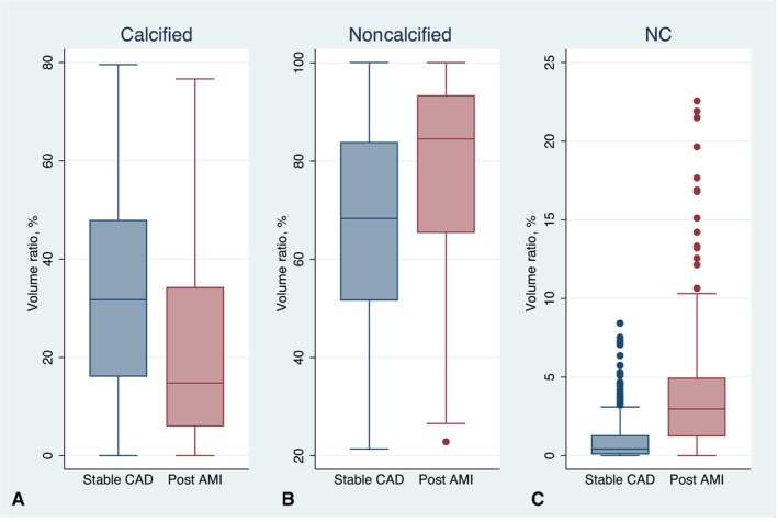
Methods and results: We analyzed 205 patients (age, 59.77±9.24 years; 92.20% men) with AMI ≤1 month and matched them with 205 patients with stable coronary artery disease (age, 60.52±10.04 years; 90.24% men) based on age, sex, and cardiovascular risk factors. Coronary computed tomography angiography scans were assessed for nonculprit plaque and vessel characteristics, plaque volumes by composition, high-risk plaques, and PVAT mean attenuation. Both patient groups exhibited similar noncalcified plaque volumes (383.35±313.23 versus 378.63±426.25 mm3, P=0.899). However, multivariable analysis revealed that patients post-AMI had a greater patient-wise noncalcified plaque volume ratio (estimate, 0.089 [95% CI, 0.053-0.125], P<0.001), largely attributed to a higher fibrofatty and necrotic core volume ratio, along with higher peri-lesion PVAT mean attenuation (estimate, 3.968 [95% CI, 2.556-5.379], P<0.001). When adjusted for vessel length, patients post-AMI had more high-risk plaques (estimate, 0.417 [95% CI, 0.298-0.536], P<0.001) per patient.

Conclusions: Patients post-AMI displayed heightened noncalcified plaque components, largely due to fibrofatty and necrotic core content, more high-risk plaques, and increased PVAT mean attenuation on a per-patient level, highlighting the necessity for refined risk assessment in patients with AMI after treatment.

Keywords: acute myocardial infarction; coronary atheroma; coronary computed tomography angiography; high‐risk plaques; residual inflammatory risk.


Figures

Figure 1
Figure 1. CONSORT (Consolidated Standards of Reporting Trials) diagram for the study.
AMI indicates acute myocardial infarction; CAD, coronary artery disease; and CCTA, coronary computed tomography angiography.
Figure 2
Figure 2. Visual representation of coronary computed tomography angiography plaque quantification.
(A) Longitudinal section of a coronary vessel with color‐coded tissue composition (red: necrotic core, yellow: fibrofatty, green: fibrous, white: calcium); (B) a necrotic core–containing cross‐section of a coronary plaque with color‐coded tissue composition (at the orange bar level of [A]); (C) a calcium‐containing cross‐section of a coronary plaque with color‐coded tissue composition (at the blue bar level of [A]); (D) cross‐section of the perivascular adipose tissue around a coronary plaque with color‐coded attenuation (at the blue bar level of [A]).
Figure 3
Figure 3. Total plaque volume (PV) and total volume of each tissue type for patients post–acute myocardial infarction (AMI; excluding culprit lesions) and patients with stable coronary artery disease (CAD).
(A) Total PV; (B) volume of calcified content; (C) volume of noncalcified content; and (D) volume of necrotic core (NC) content.
Figure 4
Figure 4. The volume ratio of each tissue type for patients post–acute myocardial infarction (AMI; excluding culprit lesions) and patients with stable coronary artery disease (CAD) after normalized to total plaque volume.
(A) Calcified content; (B) noncalcified content; (C) necrotic core (NC) content.
Figure 5
Figure 5. The regression model predicted volume ratio of each tissue type for patients with post–acute myocardial infarction (AMI; excluding culprit lesions) and patients with stable coronary artery disease (CAD) after averaging covariates.
(A) Calcified content; (B) noncalcified content; and (C) necrotic core (NC) content.
Figure 6
Figure 6. The regression model predicted per‐patient high‐risk plaques (HRPs) and high‐risk feature counts after normalized to vessel length.
AMI indicates acute myocardial infarction; CAD, coronary artery disease; LAP, low attenuation plaques; PR, positive remodeling; and SC, spotty calcification.
Figure 7
Figure 7. The regression model predicted peri‐lesion perivascular adipose tissue (PVAT) mean attenuation.
AMI indicates acute myocardial infarction; and CAD, coronary artery disease.

References

    1. Ralapanawa U, Sivakanesan R. Epidemiology and the magnitude of coronary artery disease and acute coronary syndrome: a narrative review. J Epidemiol Glob Health. 2021;11:169–177. doi: 10.2991/jegh.k.201217.001 - DOI - PMC - PubMed
    1. Motoyama S, Ito H, Sarai M, Kondo T, Kawai H, Nagahara Y, Harigaya H, Kan S, Anno H, Takahashi H, et al. Plaque characterization by coronary computed tomography angiography and the likelihood of acute coronary events in mid‐term follow‐up. J Am Coll Cardiol. 2015;66:337–346. doi: 10.1016/j.jacc.2015.05.069 - DOI - PubMed
    1. Lee SE, Sung JM, Andreini D, Al‐Mallah MH, Budoff MJ, Cademartiri F, Chinnaiyan K, Choi JH, Chun EJ, Conte E, et al. Association between changes in perivascular adipose tissue density and plaque progression. JACC Cardiovasc Imaging. 2022;15:1760–1767. doi: 10.1016/j.jcmg.2022.04.016 - DOI - PubMed
    1. Oikonomou EK, Antoniades C. The role of adipose tissue in cardiovascular health and disease. Nat Rev Cardiol. 2019;16:83–99. doi: 10.1038/s41569-018-0097-6 - DOI - PubMed
    1. Montone RA, Niccoli G, Crea F, Jang IK. Management of non‐culprit coronary plaques in patients with acute coronary syndrome. Eur Heart J. 2020;41:3579–3586. doi: 10.1093/eurheartj/ehaa481 - DOI - PubMed

Publication types

MeSH terms

LinkOut - more resources