. 2024 Nov 19;98(11):e0119424.
doi: 10.1128/jvi.01194-24. Epub 2024 Oct 29.

Structures of Epstein-Barr virus and Kaposi's sarcoma-associated herpesvirus virions reveal species-specific tegument and envelope features

Affiliations

Structures of Epstein-Barr virus and Kaposi's sarcoma-associated herpesvirus virions reveal species-specific tegument and envelope features

James Zhen et al. J Virol. .

Abstract

Epstein-Barr virus (EBV) and Kaposi's sarcoma-associated herpesvirus (KSHV) are classified into the gammaherpesvirus subfamily of Herpesviridae, which stands out from its alpha- and betaherpesvirus relatives due to the tumorigenicity of its members. Although structures of human alpha- and betaherpesviruses by cryogenic electron tomography (cryoET) have been reported, reconstructions of intact human gammaherpesvirus virions remain elusive. Here, we structurally characterize extracellular virions of EBV and KSHV by deep learning-enhanced cryoET, resolving both previously known monomorphic capsid structures and previously unknown pleomorphic features beyond the capsid. Through subtomogram averaging and subsequent tomogram-guided sub-particle reconstruction, we determined the orientation of KSHV nucleocapsids from mature virions with respect to the portal to provide spatial context for the tegument within the virion. Both EBV and KSHV have an eccentric capsid position and polarized distribution of tegument. Tegument species span from the capsid to the envelope and may serve as scaffolds for tegumentation and envelopment. The envelopes of EBV and KSHV are less densely populated with glycoproteins than those of herpes simplex virus 1 (HSV-1) and human cytomegalovirus (HCMV), representative members of alpha- and betaherpesviruses, respectively. Also, we observed fusion protein gB trimers exist within triplet arrangements in addition to standalone complexes, which is relevant to understanding dynamic processes such as fusion pore formation. Taken together, this study reveals nuanced yet important differences in the tegument and envelope architectures among human herpesviruses and provides insights into their varied cell tropism and infection.

Importance: Discovered in 1964, Epstein-Barr virus (EBV) is the first identified human oncogenic virus and the founding member of the gammaherpesvirus subfamily. In 1994, another cancer-causing virus was discovered in lesions of AIDS patients and later named Kaposi's sarcoma-associated herpesvirus (KSHV), the second human gammaherpesvirus. Despite the historical importance of EBV and KSHV, technical difficulties with isolating large quantities of these viruses and the pleiomorphic nature of their envelope and tegument layers have limited structural characterization of their virions. In this study, we employed the latest technologies in cryogenic electron microscopy (cryoEM) and tomography (cryoET) supplemented with an artificial intelligence-powered data processing software package to reconstruct 3D structures of the EBV and KSHV virions. We uncovered unique properties of the envelope glycoproteins and tegument layers of both EBV and KSHV. Comparison of these features with their non-tumorigenic counterparts provides insights into their relevance during infection.

Keywords: Epstein-Barr virus; Kaposi's sarcoma-associated herpesvirus; electron microscopy; glycoproteins; herpesviruses; human herpesviruses; virion structure.


Conflict of interest statement

The authors declare no conflict of interest.

Figures

Fig 1
Fig 1
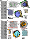
Isolated EBV and KSHV virion samples. (a) CryoEM screening image of isolated EBV virion sample. Intact EBV virion is indicated by arrow (orchid). Scale bar, 200 nm. (b) Low-dose image of EBV virion sample with 10-nm gold fiducials at a tilt angle of zero degrees for tomography. Intact EBV virion is indicated by arrow (orchid). Scale bar, 200 nm. (c) CryoEM screening image of isolated KSHV virion sample. Example of intact KSHV virions is indicated by arrow (sky). Scale bar, 200 nm. (d) Low-dose image of KSHV virion sample with 5-nm gold fiducials at a tilt angle of zero degrees for tomography. Example of intact KSHV virions is indicated by arrow (sky). Red arrow indicates broken virion. Scale bar, 200 nm.
Fig 2
Fig 2
CryoET of EBV and KSHV. (a, e) Series of tomogram slices from weighted back-projection reconstruction with SIRT-like filter along the z-axis of virion. Numbers correspond to location in surface rendering depiction (c, g). Scale bar, 80 nm. (b, f) Series of tomogram slices from isotropic reconstruction along the z-axis of virion. EM density at top and bottom represents envelope, which indicates intact virion. Scale bar, 80 nm. (c, g) Surface renderings of virion reconstructions were cropped at the top and bottom poles (red lines) prior to segmentation to improve interpretability. Numbers indicate locations of corresponding tomogram slices (b, f). Scale bar, 40 nm. (d, h) Sectional view of 3D surface rendering of virion colored by different components. Tomogram slices of corresponding virion component densities are indicated by their respective colors. Scale bar, 40 nm.
Fig 3
Fig 3
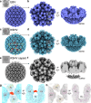
Subtomogram average reconstructions of EBV and KSHV capsid. (a, c) Subtomogram average of virion capsid with icosahedral symmetry imposed. Top left shows a tomogram slice of representative intact virion used for capsid reconstruction. In KSHV, additional densities of unknown identity are indicated (red circles). Twofold and threefold symmetry axes are represented by orange lines and yellow triangle, respectively. Triplex nomenclature is annotated. (b, d) Orthogonal views of tomogram-guided sub-particle reconstruction of capsid vertex with C5 symmetry imposed. Hexon, triplex, fivefold vertex, and dsDNA are labeled. (e) CryoEM map of naked KSHV capsid, devoid of tegument with icosahedral symmetry imposed, was low-pass filtered to the same resolution as KSHV subtomogram average. Top left shows a cryoEM image of representative naked KSHV capsid devoid of tegument used for reconstruction. (f) Orthogonal views of capsid vertex density cropped from naked KSHV capsid reconstruction. (g) Overhead and side view of unfilled KSHV density from (c) indicated in red. Atomic models of neighboring triplexes (Tb, Te) and MCP/SCP (E2, C5) (PDB ID: 6B43) are fitted around the unfilled density. (h) Overhead and side view of region shown in (g) for naked KSHV capsid.
Fig 4
Fig 4
Tomogram-guided sub-particle reconstruction of KSHV penton and portal vertices. (a) Tomogram-guided sub-particle reconstruction of KSHV penton vertex with C5 symmetry (light blue). (b) Tomogram-guided sub-particle reconstruction of KSHV portal vertex with C5 symmetry (turquoise). (c) Side projection of KSHV penton vertex. (d) Side projection of KSHV portal vertex. Portal cap and portal complex are indicated. (e) Secondary structure fit of KSHV MCP and SCP atomic model (PDB ID: 6B43) into cryoEM map of KSHV penton vertex (left) and KSHV portal vertex (right). (f) Segmentation of CVSC density (teal) in KSHV portal vertex. Triplex, MCP, and SCP are colored orange, purple, and pink, respectively. (g) Two classes of KSHV portal vertex (green, blue). Inset shows region of portal cap density with portal cap and binding sites of CVSC indicated by red dotted lines. (h) Overlay of portal cap and CVSC binding site schematic from (g), adjusted for rotation of portal cap. CVSC binding sites are colored by the color of the respective class (green, blue). The angles between the portal cap and the connecting density to CVSC are indicated.
Fig 5
Fig 5
KSHV tegument in context of the portal and envelope. (a) The location and orientation of KSHV capsid 3D reconstruction (sky) mapped back into a tomogram. Portal vertex (red) and penton vertices (pink) for each virus are indicated. (b) Globular densities in tegument (orange) located near the portal (red) for two representative virions. (c) Circumnavigation of envelope by discontinuous density resembling a dotted line (between two orange lines). (d) Long, thin densities in outer tegument bordering envelope. (e) Outer tegument (orange) associated with envelope protein (red) endodomains at a protrusion with few tegument densities elsewhere. (f) Long, thin densities spanning from capsid to envelope. Portal vertex (red) and adjacent penton vertices (pink) are indicated. Spanning densities are present in both presence (left) and absence (right) of tegument bulk. Scale bars, 40 nm.
Fig 6
Fig 6
Envelope protein organization. (a) Comparison of tomogram slices between EBV, KSHV, HSV-1, and HCMV. Clusters of envelope proteins are indicated by red arrows. Scale bar, 40 nm. (b) Count of envelope proteins per virion for indicated herpesvirus species. Statistical analysis used ordinary one-way analysis of variance (ANOVA) with Tukey’s test for multiple comparisons (*, P < 0.05). (c) Subtomogram average reconstruction of large envelope proteins from clusters in KSHV. Postfusion gB atomic model (blue; PDB ID: 3FVC) fits the cryoET density.

Update of

References

    1. Schiller JT, Lowy DR. 2021. An introduction to virus infections and human cancer. Recent Results Cancer Res 217:1–11. doi:10.1007/978-3-030-57362-1_1 - DOI - PMC - PubMed
    1. Wen KW, Wang L, Menke JR, Damania B. 2022. Cancers associated with human gammaherpesviruses. FEBS J 289:7631–7669. doi:10.1111/febs.16206 - DOI - PMC - PubMed
    1. Bjornevik K, Cortese M, Healy BC, Kuhle J, Mina MJ, Leng Y, Elledge SJ, Niebuhr DW, Scher AI, Munger KL, Ascherio A. 2022. Longitudinal analysis reveals high prevalence of Epstein-Barr virus associated with multiple sclerosis. Science 375:296–301. doi:10.1126/science.abj8222 - DOI - PubMed
    1. Chang Y, Cesarman E, Pessin MS, Lee F, Culpepper J, Knowles DM, Moore PS. 1994. Identification of herpesvirus-like DNA sequences in AIDS-associated Kaposi’s sarcoma. Science 266:1865–1869. doi:10.1126/science.7997879 - DOI - PubMed
    1. Cesarman E, Damania B, Krown SE, Martin J, Bower M, Whitby D. 2019. Kaposi sarcoma. Nat Rev Dis Primers 5:9. doi:10.1038/s41572-019-0060-9 - DOI - PMC - PubMed

LinkOut - more resources