. 2024 Jan-Dec:33:9636897241291278.
doi: 10.1177/09636897241291278.

Exogenous Mitochondrial Transplantation Facilitates the Recovery of Autologous Nerve Grafting in Repairing Nerve Defects

Affiliations

Exogenous Mitochondrial Transplantation Facilitates the Recovery of Autologous Nerve Grafting in Repairing Nerve Defects

Dongdong Li et al. Cell Transplant. 2024 Jan-Dec.

Abstract

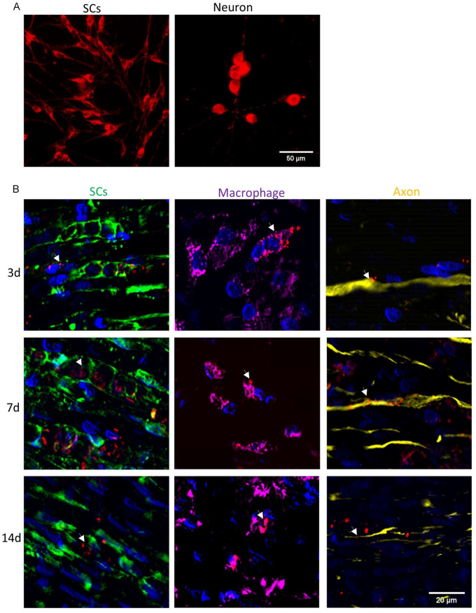
Autologous nerve transplantation (ANT) remains the gold standard for treating nerve defects. However, its efficacy in nerve repair still requires improvement. Mitochondrial dysfunction resulting from nerve injury may be a significant factor limiting nerve function restoration. This study investigated the impact of supplementing exogenous mitochondria (EM) in ANT and explored its effect on the efficacy of ANT in nerve repair. SD rats were used to prepare a model of a 10 mm sciatic nerve defect repaired by ANT (Auto group) and a model of ANT supplemented with EM (Mito group). At 12 weeks post-operation, functional, neurophysiological, and histological evaluations of the target organ revealed that the Mito group exhibited significantly better outcomes compared with the Auto group, with statistically significant differences (P < 0.05). In vitro experiments demonstrated that EM could be endocytosed by Schwann cells (SCs) and dorsal root ganglion neurons (DRGs) when co-cultured. After endocytosis by SCs, immunofluorescence staining of autophagy marker LC3II and mitochondrial marker Tomm20, as well as adenoviral fluorescence labeling of lysosomes and mitochondria, revealed that EM could promote autophagy in SCs. CCK8 and EDU assays also indicated that EM significantly promoted SCs proliferation and viability. After endocytosis by DRGs, EM could accelerate axonal growth rate. A sciatic nerve defect repair model prepared using Thy1-YFP-16 mice also revealed that EM could accelerate axonal growth in vivo, with statistically significant results (P < 0.05). This study suggests that EM enhances autophagy in SCs, promotes SCs proliferation and viability, and increases the axonal growth rate, thereby improving the efficacy of ANT. This research provides a novel therapeutic strategy for enhancing the efficacy of ANT in nerve repair.

Keywords: Schwann cells; autologous nerve transplantation; functional recovery; mitochondria transplantation; nerve defect.


Conflict of interest statement

Declaration of Conflicting InterestsThe author(s) declared no potential conflicts of interest with respect to the research, authorship, and/or publication of this article.

Figures

Graphical Abstract
Graphical Abstract
Figure 1.
Figure 1.
(A) Footprints of rats at 12 weeks post-surgery, with LH representing the control side and RH representing the experimental side. (B) Contact area between rat toes and the ground surface at 12 weeks post-surgery. (C) Statistical analysis of sciatic nerve function index at 4, 8, 12 weeks post-surgery. Data are expressed as mean ± SD.
Figure 2.
Figure 2.
(A) Toluidine blue staining of regenerated myelinated nerve fibers. (B) TEM scanning of regenerated nerve fibers. (C) Staining of regenerated nerve fibers’ axons and SCs, with green representing axons and red representing SCs. (D) Statistical analysis of myelin density. (E) Statistical analysis of myelin thickness. (F) Statistical analysis of axon fluorescence density. (G) Statistical analysis of SCs fluorescence density. Data are expressed as mean ± SD.
Figure 3.
Figure 3.
(A) Gross images of the gastrocnemius muscle at 3 months post-surgery. (B) Masson staining of cross-sections of the gastrocnemius muscle. (C) Compound muscle action potential waveforms of the gastrocnemius muscle. (D) Statistical analysis of wet weight recovery rates of the gastrocnemius muscle. (E) Statistical analysis of cross-sectional areas of muscle fibers in the gastrocnemius muscle. (F) Statistical analysis of CMAP recovery rates of the gastrocnemius muscle. Data are expressed as mean ± SD.
Figure 4.
Figure 4.
Endocytosis of EM. (A) Fluorescence microscopy images showing abundant red fluorescence within the cytoplasm of both neuron and SCs, indicating the presence of exogenous mitochondria. (B) Fluorescence microscopy images of autologous nerve grafts injected with exogenous mitochondria at 3 days post-surgery. The images demonstrate the endocytosis of exogenous mitochondria by SCs (green), macrophages (purple), and axons (yellow). The red fluorescence represents exogenous mitochondria. Fluorescence signals persisted at 14 days post-surgery.
Figure 5.
Figure 5.
SCs Proliferation Experiment. (A) EdU staining images of SCs, with red representing newly proliferated cells and blue representing all cells. (B) Ratio of double-stained cells (EdU-positive) to the total number of cells. (C) Absorbance values of SCs after CCK8 test at different time points. Data are expressed as mean ± SD.
Figure 6.
Figure 6.
Axonal Growth Length. (A) NF-200 and S100 staining images of DRG and SCs in vitro. Axons are represented in green, while SCs are represented in red. (B) Process of axonal growth in vivo. (C) Statistical analysis of axonal length after 7 days of in vitro culture. (D) Statistical analysis of axonal length after 10 days of neural defect repair in vivo. Data are expressed as mean ± SD.
Figure 7.
Figure 7.
Induction of autophagy in SCs by exogenous mitochondria. (A) Immunofluorescence staining of the mitochondrial marker protein TOMM20 in SCs. Cell nuclei are shown in blue, and TOMM20 is depicted in red. (B) Immunofluorescence staining of the lysosomal marker protein LC3B in SCs. Cell nuclei are shown in blue, and LC3B is depicted in red. (C) Fluorescence images of SCs transfected with adenovirus carrying LC3-GFP and mito-keima, labeling lysosomal LC3 and mitochondrial keima fluorescent proteins, respectively. After addition of exogenous mitochondria, enhancement of mitochondrial autophagy and increased lysosomal expression are observed in SCs. (D) Statistical analysis of fluorescence intensity after LC3 labeling of lysosomes. (E) Statistical analysis of autophagy index after keima labeling of mitochondria. (F) Statistical analysis of TOMM20 immunofluorescence intensity. (G) Statistical analysis of LC3 immunofluorescence intensity. Data are expressed as mean ± SD.

Similar articles

Cited by

References

    1. Namini MS, Daneshimehr F, Beheshtizadeh N, Mansouri V, Ai J, Jahromi HK, Ebrahimi-Barough S. Cell-free therapy based on extracellular vesicles: a promising therapeutic strategy for peripheral nerve injury. Stem Cell Res Ther. 2023;14(1):254. - PMC - PubMed
    1. Liu X, Zou D, Hu Y, He Y, Lu J. Research progress of low-intensity pulsed ultrasound in the repair of peripheral nerve injury. Tissue Eng Part B Rev. 2023;29(4):414–28. - PubMed
    1. Kim J, Jeon J, Lee JY, Khoroldulam B, Choi SG, Bae JY, Hyun JK, Kang SK. Electroceuticals for regeneration of long nerve gap using biodegradable conductive conduits and implantable wireless stimulator. Adv Sci (Weinh). 2023;10(24):e2302632. - PMC - PubMed
    1. Guan Y, Ren Z, Yang B, Xu W, Wu W, Li X, Zhang T, Li D, Chen S, Bai J, Song X, et al.. Dual-bionic regenerative microenvironment for peripheral nerve repair. Bioact Mater. 2023;26:370–86. - PMC - PubMed
    1. Wang B, Lu CF, Liu ZY, Han S, Wei P, Zhang DY, Kou YH, Jiang BG. Chitin scaffold combined with autologous small nerve repairs sciatic nerve defects. Neural Regen Res. 2022;17(5):1106–14. - PMC - PubMed

LinkOut - more resources