. 2024 Oct 29;22(10):e3002884.
doi: 10.1371/journal.pbio.3002884. eCollection 2024 Oct.

The future of transcranial ultrasound as a precision brain interface

Affiliations

The future of transcranial ultrasound as a precision brain interface

Keith Murphy et al. PLoS Biol. .

Abstract

Our understanding of brain circuit operations and disorders has rapidly outpaced our ability to intervene and restore them. Developing technologies that can precisely interface with any brain region and circuit may combine diagnostics with therapeutic intervention, expediting personalised brain medicine. Transcranial ultrasound stimulation (TUS) is a promising noninvasive solution to this challenge, offering focal precision and scalability. By exploiting the biomechanics of pressure waves on brain tissue, TUS enables multi-site targeted neuromodulation across distributed circuits in the cortex and deeper areas alike. In this Essay, we explore the emergent evidence that TUS can functionally test and modify dysfunctional regions, effectively serving as a search and rescue tool for the brain. We define the challenges and opportunities faced by TUS as it moves towards greater target precision and integration with advanced brain monitoring and interventional technology. Finally, we propose a roadmap for the evolution of TUS as it progresses from a research tool to a clinically validated therapeutic for brain disorders.


Conflict of interest statement

We have read the journal’s policy and the author K.M. of this manuscript has the following competing interests: K.M. is a co-founder and shareholder of Attune Neuroscience.

Figures

Fig 1
Fig 1. Focused ultrasound device selection and stimulation planning.
Graphic illustrations of (a) an ultrasound array hemisphere, (b) 2 multielement transducers, and (c) a single-element transducer. (d) MRI capture of a brain anatomy through T1 imaging and skull density approximation through ultrashort echo time imaging. (e) Various stages of image segmentation prior to (f) acoustic simulation of waveform focusing in cranial space. MRI, magnetic resonance imaging.
Fig 2
Fig 2. TUS in precision neuroscience: a search and rescue tool.
(a) Participant in an MRI scanner performs a task. (b) Inferential level where brain mapping is made between cognitive processes and mechanistic features of brain regions function. (c–e) Causal manipulation can happen at the region level (c), allowing double dissociation intervention, (d) at the circuit level, allowing disconnection logic and the impact or circuit function or (e) at the subregion level, including subdivisions of anatomically defined brain nuclei. (f) Ontological levels relevant to TUS, defining the relationship between neural mechanisms (neural level) to inferences and modelling of brain target and circuits theoretical properties (inference level) to clinical interventional (clinical level). Top panel: At the neural level, a brain circuit mediates different cognitive processes (triggered exogenously or endogenously), denoted with Greek letters, within defined regions coupled with others. Middle panel: At the inferential level, theoretical models attempt to infer the relationship between behaviours and brain regions. This can be refined by causally manipulating brain regions with TUS to estimate the impact of brain perturbation on certain hypothesised cognitive processes. Bottom panel: Once an area is identified to relate to a specific cognitive process mapped onto psychiatric traits, this brain region can be perturbed with TUS to restore healthy functioning. MRI, magnetic resonance imaging; TUS, transcranial ultrasound stimulation.
Fig 3
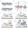
Fig 3. Conceptualizations of next generation TUS innovations.
(a) Cell-type biased neuromodulation through application of variant TUS waveforms. (b) Illustration of BMI electrodes recording local field potentials during TUS exposure. Note the potential mechanical “artefact” recorded during the TUS delivery window which may be used to validate that the target is being exposed to ultrasound. (c) Illustration of lipid rafts within the neuronal membrane. Comparison of lipid raft and membrane disruption with TUS only (left), compared to TUS with microbubbles which expand during the low-pressure phase. Microbubbles may enhance mechanically derived neuromodulatory effects. BMI, brain–machine interface; TUS, transcranial ultrasound stimulation.
Fig 4
Fig 4. Circuit tracing as a method of cell type selective neuromodulation.
A graphical illustration of 3 partially overlapping cell types shows how a focus can be moved along the tracts of a target neuronal circuit (purple). While cell bodies of off target populations may partially overlap at the targets cell bodies (blue) or axonal tracts (orange), exposure is limited to foci at a subset of raster positions. In this hypothetical scenario, only the purple cell type experiences repeated TUS induced spiking increases over the stimulation period. TUS, transcranial ultrasound stimulation.
Fig 5
Fig 5. Conceptualisation of next generation TUS interventions.
Clinical and at-home intervention are shown and involve meticulous verification of target engagement to ensure precision and safety. In-clinic use may include MRI to plan the intervention. Advances in simulation accuracy and quality control research will establish the reliability of TUS procedures. At-home TUS systems could be used for chronic symptom alleviation but require personalised MRI guidance and regulatory approval to ensure safety and efficacy. MRI, magnetic resonance imaging; TUS, transcranial ultrasound stimulation.

Similar articles

Cited by

References

    1. Silberberg D, Anand NP, Michels K, Kalaria RN. Brain and other nervous system disorders across the lifespan—global challenges and opportunities. Nature. 2015;527:S151–154. doi: 10.1038/nature16028 - DOI - PubMed
    1. Harmsen IE, Elias GJB, Beyn ME, Boutet A, Pancholi A, Germann J, et al.. Clinical trials for deep brain stimulation: Current state of affairs. Brain Stimul. 2020;13:378–385. doi: 10.1016/j.brs.2019.11.008 - DOI - PubMed
    1. Feinberg DA, Beckett AJS, Vu AT, Stockmann J, Huber L, Ma S, et al.. Next-generation MRI scanner designed for ultra-high-resolution human brain imaging at 7 Tesla. Nat Methods. 2023;20:2048–2057. doi: 10.1038/s41592-023-02068-7 - DOI - PMC - PubMed
    1. Myszczynska MA, Ojamies PN, Lacoste AMB, Neil D, Saffari A, Mead R, et al.. Applications of machine learning to diagnosis and treatment of neurodegenerative diseases. Nat Rev Neurol. 2020;16:440–456. doi: 10.1038/s41582-020-0377-8 - DOI - PubMed
    1. Ma D, Gulani V, Seiberlich N, Liu K, Sunshine JL, Duerk JL, et al.. Magnetic resonance fingerprinting. Nature. 2013;495:187–192. doi: 10.1038/nature11971 - DOI - PMC - PubMed

LinkOut - more resources