. 2024 Oct 29;20(10):e1012660.
doi: 10.1371/journal.ppat.1012660. eCollection 2024 Oct.

BiP/GRP78 is a pro-viral factor for diverse dsDNA viruses that promotes the survival and proliferation of cells upon KSHV infection

Affiliations

BiP/GRP78 is a pro-viral factor for diverse dsDNA viruses that promotes the survival and proliferation of cells upon KSHV infection

Guillermo Najarro et al. PLoS Pathog. .

Abstract

The Endoplasmic Reticulum (ER)-resident HSP70 chaperone BiP (HSPA5) plays a crucial role in maintaining and restoring protein folding homeostasis in the ER. BiP's function is often dysregulated in cancer and virus-infected cells, conferring pro-oncogenic and pro-viral advantages. We explored BiP's functions during infection by the Kaposi's sarcoma-associated herpesvirus (KSHV), an oncogenic gamma-herpesvirus associated with cancers of immunocompromised patients. Our findings reveal that BiP protein levels are upregulated in infected epithelial cells during the lytic phase of KSHV infection. This upregulation occurs independently of the unfolded protein response (UPR), a major signaling pathway that regulates BiP availability. Genetic and pharmacological inhibition of BiP halts KSHV viral replication and reduces the proliferation and survival of KSHV-infected cells. Notably, inhibition of BiP limits the spread of other alpha- and beta-herpesviruses and poxviruses with minimal toxicity for normal cells. Our work suggests that BiP is a potential target for developing broad-spectrum antiviral therapies against double-stranded DNA viruses and a promising candidate for therapeutic intervention in KSHV-related malignancies.


Conflict of interest statement

The authors have declared that no competing interests exist.

Figures
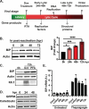
Fig 1
Fig 1. BiP is upregulated during the KSHV lytic cycle.
(A) Schematic of lytic reactivation in iSLK.219 cells (B) BiP is upregulated at the protein level in a time course of reactivation. (Left) iSLK.219 cells were treated with Dox (1 μg/ml) to induce RTA expression and viral reactivation. Whole-cell lysates collected at the indicated times were analyzed by immunoblot. Actin: loading control. (Right) Image densitometry quantification of the immunoblot (C) BiP upregulation is independent of late viral gene expression. Viral DNA replication was inhibited in iSLK.219 cells by pretreatment with PFA (100 nM) for 24h before induction with Dox. Whole-cell lysates collected at the indicated times were analyzed by immunoblot. Actin: loading control. (D) Immunoblot of GRP94, calreticulin, and actin during KSHV reactivation in iSLK.219 cells (E) BiP upregulation is post-transcriptional. qRT-PCR quantification of BiP mRNA in a time course of reactivation in iSLK.219 untreated or treated with Tg (100nM) for 4h. Note the high levels of BiP mRNA in cells undergoing acute ER stress. N = 3 independent biological replicates for (B, C, D, and E). Values in (B, E) are average ±SEM. Statistical significance in (B) was calculated using a one-way ANOVA (*P = 0.01, **P = 0.004, **P = <0.0001).
Fig 2
Fig 2. BiP is post-transcriptionally upregulated independently of ATF6 and XBP1.
(A-C) IRE1 is phosphorylated in lytic iSLK.219 cells without detectable XBP1 splicing. Cells were reactivated by treatment with Dox (1 μg/ml). At the indicated times, the cells were treated with Tg (100 nM) for 4h to induce acute ER stress. (A) Whole-cell lysates were collected and analyzed by immunoblot for total (IRE1) or phosphorylated IRE1 (IRE1-P), spliced XBP1 (XBP1s), and actin (loading control). (B) RT-PCR detection of unspliced (u) and spliced (s) XBP1 mRNA. (C) Image densitometry quantification of the data in (B). (D-H) XBP1 and ATF6 are not required for BiP protein upregulation or infectious virus production during the KSHV lytic cycle. (D) CRISPRi-based knockdown of XBP1 (XBP1-KD) in iSLK.219-dCas9 cells. Cells (NS and XBP1-KD) were induced with Dox (1 μg/ml) for 24h. Cells were treated with Tg (100 nM) for 4h before collection. Whole-cell lysates were analyzed by immunoblot. Actin: loading control. (E) The supernatants of cells treated as in (D) were collected and used to spinoculate uninfected iSLK cells. The percent of GFP expression was determined by automated cell counting and used as a proxy for infectious virus levels in the supernatants. (F) iSLK.219 cells were treated with the ATF6 inhibitor CeapinA7 (6 μM) for 2h before induction with Dox (1 μg/ml). Whole-cell lysates were collected at the indicated times and analyzed by immunoblot. (G) CRISPRi-based knockdown of ATF6 (ATF6KD) in iSLK.219-dCas9 cells. ATF6-KD cells were treated as in (D). The knockdown of ATF6 expression was determined by qRT-PCR. (H) Supernatants from ATF6-KD cells were collected and processed as described in (E). N = 3 independent biological replicates. Values in (C, E, F) are average ±SEM. Statistical significance was calculated using a one-way ANOVA (*P = 0.01) in (C) or a paired t-test (E and H).
Fig 3
Fig 3. BiP is a pro-viral factor in KSHV-infected cells.
(A-B) BiP inhibition with HA15 disrupts the lytic cycle iSLK.219s. (A) Cells were treated with HA15 (10 μM) 24h before reactivation with Dox (1 μg/ml). At the indicated times, whole-cell lysates were collected and analyzed by immunoblot for viral proteins (Immediate early: KbZip-nuclear, ORF57-nuclear, Early: ORF45-nuclear/cytosolic, Late: K8.1-glycoprotein). Actin: loading control. (B) Supernatants from iSLK.219 cells treated with HA15 were collected at 72h post reactivation and used to infect naïve iSLK cells. GFP expression was determined by automated cell counting at 48h post-infection and used as a proxy for virus production. (C-D) Silencing of BiP reduces viral reactivation and infectious virion production. (C) iSLK.219 cells were reactivated with Dox, following BiP siRNA-mediated silencing for 48h. Lysates were collected at 72h post-reactivation and analyzed by immunoblot for viral factors. siRNA–untransfected, NT non-targeting (D) Supernatants from BiP-KD cells treated as in (C) were collected and processed as described in (B). (E-F) Inhibition of BiP blocks the lytic cycle in TREx-BCBL-1-RTA cells. (E) Cells were treated with HA15 (10 μM) for 24h before induction with Dox (1 μg/ml). At 48h post-infection, whole cell lysates were collected and analyzed by immunoblot. Actin: loading control. (F) Total DNA was isolated from cells treated as in (E), and viral DNA was quantified by qRT-PCR. N = 3 independent biological replicates. Values in (B, D, F) are average ±SEM. Statistical significance was calculated using a paired t-test (B and D) (*P = 0.01, **P = 0.002) or a two-way ANOVA (F) (** P = 0.0065).
Fig 4
Fig 4. BiP inhibition disrupts the KSHV lytic cycle.
(A-C) Total RNA was isolated from latent and lytic iSLK.219 cells in the presence or absence of HA15. RNAseq libraries were prepared, sequenced, and aligned to the KSHV genome. (A) Pairwise comparison of the RNA-seq samples generated in this study. (B) Boxplot of the Log2 of normalized counts of KSHV genes in latent and lytic iSLK.219 cells at 72h post-reactivation in the presence or absence of HA15. (C) Heatmap of the scaled normalized counts (Z score) for all KSHV genes ordered by genomic position in lytic iSLK.219 cells ± HA15.
Fig 5
Fig 5. HA15 causes strong cytostasis in latent PEL-derived cells and cytotoxicity in KSHV-infected LEC.
(A-B) HA15 treatment differentially reduces cell numbers compared to cell viability in TREx-BCBL-1 cells. Latent TREx-BCBL-1 cells were treated with increasing concentrations of HA15 (0–50 μM) for 72h. The total number of viable cells (A) and the percent of dead TREx-BCBL-1 cells (B) were determined by automated cell counting following trypan blue staining. (C-D) HA15 treatment does not cause cytostasis nor cytotoxicity in primary B cells. Primary Peripheral B-cells were treated with increasing concentrations of HA15 (0–50 μM) for 72h. The total number of viable cells (C) and the percent of live cells (D) were determined as described in (A-B). N = 3 independent biological replicates. Values are average ±SEM. (E-G) Primary Lymphatic endothelial cells were infected with KSHV.219 and selected with puromycin for 7–14 days. (A) Whole-cell lysates from uninfected (LEC) or infected (KLECs) were collected and analyzed by immunoblot. Actin: loading control. (B-C) LECs and KLECs were treated with HA15 (10 μM) for 72h. Cell viability was evaluated by ATP quantification using CellTiter-Glo (B) and microscopy at 0h and 72h post-treatment (C.).
Fig 6
Fig 6. The BiP inhibitor HA15 has a broad-spectrum antiviral effect on herpesviruses and poxviruses.
(A-B) Primary human fibroblasts (NHDF) were Infected at a low multiplicity of infection (MOI; HSV-1 at MOI 0.001, HCMV at MOI 0.1, and VV at MOI 0.01) in the presence or absence of HA15 (10 μM or 30 μM). (A) The spread of infection was determined at different times post-infection by measuring the expression of virus-encoded GFP in HSV-1-GFP and HCMV-GFP infected cells or by immunofluorescence using a polyclonal antibody against Vaccinia virus. (B) Representative images of the samples measured in (A). (C) The effect of HA15 treatment on the viability of NDHF (1 or 6 days) and iSLK.219s (3 days) was evaluated by measuring LDH release. N = 3 (A), N = 6 (B) independent biological replicates. Values are average ±SEM.

References

    1. Stern-Ginossar N, Thompson SR, Mathews MB, Mohr I. Translational Control in Virus-Infected Cells. Cold Spring Harb Perspect Biol. 2019;11: a033001. doi: 10.1101/cshperspect.a033001 - DOI - PMC - PubMed
    1. Aviner R, Frydman J. Proteostasis in Viral Infection: Unfolding the Complex Virus-Chaperone Interplay. Cold Spring Harb Perspect Biol. 2020;12. doi: 10.1101/cshperspect.a034090 - DOI - PMC - PubMed
    1. Paladino L, Vitale AM, Bavisotto CC, de Macario EC, Cappello F, Macario AJL, et al.. The role of molecular chaperones in virus infection and implications for understanding and treating covid-19. Journal of Clinical Medicine. MDPI; 2020. pp. 1–15. doi: 10.3390/jcm9113518 - DOI - PMC - PubMed
    1. Zhang X, Yu W. Heat shock proteins and viral infection. Front Immunol. 2022;13. doi: 10.3389/fimmu.2022.947789 - DOI - PMC - PubMed
    1. Evelyne K, Sébastien C, Valentin B, Laurence D, Natalia B-B, Oleg D, et al.. Endoplasmic Reticulum Chaperones in Viral Infection: Therapeutic Perspectives. Microbiology and Molecular Biology Reviews. 2021;85: e00035–21. doi: 10.1128/MMBR.00035-21 - DOI - PMC - PubMed

Substances