. 2024 Nov 5;121(45):e2412358121.
doi: 10.1073/pnas.2412358121. Epub 2024 Oct 29.

A molecular view of peptoid-induced acceleration of calcite growth

Affiliations

A molecular view of peptoid-induced acceleration of calcite growth

Mingyi Zhang et al. Proc Natl Acad Sci U S A. .

Abstract

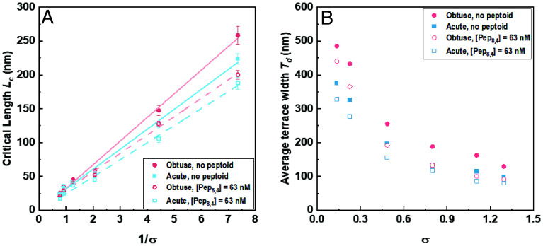
The extensive deposits of calcium carbonate (CaCO3) generated by marine organisms constitute the largest and oldest carbon dioxide (CO2) reservoir. These organisms utilize macromolecules like peptides and proteins to facilitate the nucleation and growth of carbonate minerals, serving as an effective method for CO2 sequestration. However, the precise mechanisms behind this process remain elusive. In this study, we report the use of sequence-defined peptoids, a class of peptidomimetics, to achieve the accelerated calcite step growth kinetics with the molecular level mechanistic understanding. By designing peptoids with hydrophilic and hydrophobic blocks, we systematically investigated the acceleration in step growth rate of calcite crystals using in situ atomic force microscopy (AFM), varying peptoid sequences and concentrations, CaCO3 supersaturations, and the ratio of Ca2+/ HCO3-. Mechanistic studies using NMR, three-dimensional fast force mapping (3D FFM), and isothermal titration calorimetry (ITC) were conducted to reveal the interactions of peptoids with Ca2+ and HCO3- ions in solution, as well as the effect of peptoids on solvation and energetics of calcite crystal surface. Our results indicate the multiple roles of peptoid in facilitating HCO3- deprotonation, Ca2+ desolvation, and the disruption of interfacial hydration layers of the calcite surface, which collectively contribute to a peptoid-induced acceleration of calcite growth. These findings provide guidelines for future design of sequence-specific biomimetic polymers as crystallization promoters, offering potential applications in environmental remediation (such as CO2 sequestration), biomedical engineering, and energy storage where fast crystallization is preferred.

Keywords: CO2 sequestration; atomic force microscopy; biomimetic polymer; crystal growth.


Conflict of interest statement

Competing interests statement:The authors declare no competing interest.

Figures

Fig. 1.
Fig. 1.
(A) Sequences of peptoids employed to regulate calcite growth kinetics. (BE) in situ AFM images showing the morphology of a calcite growth hillock in a supersaturated solution (σ = 0.226) for Pep8,4 concentrations of (B) 0, (C) 31.6 nM, (D) 63.1 nM, and (E) 158.5 nM. (F) Measured step speeds as a function of concentration for all peptoids having four Npe groups (NcenNpe4, where n = 4, 6, 8, 10, 12). (G) Measured step speeds as a function of concentration for all peptoids having eight Nce groups (Nce8Npem, where m = 3, 4, 7). (H) Correlation between the optimal peptoid concentration and the values of n and m. (I) Correlation between the maximum enhancement ratio and the values of n and m.
Fig. 2.
Fig. 2.
(A and B) Acceleration of calcite obtuse (A) and acute (B) step rate as a function of Pep8,4 concentration at different solution supersaturations (σcalcite = 0.136, 0.226, 0.486, 0.799, 1.294), where v and v0 denote step rates with and without peptoids, respectively. The curves are presented using a logarithmic scale for better clarity. (C and D) The relationship between step speeds and the concentration of Ca2+ for both obtuse (C) and acute (D) steps at different concentrations of Pep8,4. (E and F) The relationship between step speeds and activity ratios for both obtuse (E) and acute (F) steps at a constant supersaturation and pH (σcalcite = 0.226, pH = 8.25) at varied concentration of Pep8,4. Error bars are one SD.
Fig. 3.
Fig. 3.
(A) Dependence of critical length of calcite steps on inverse supersaturation (1/σ) in the absence and presence of Pep8,4 (63.1 nM). The bars represent SD. (B) Dependence of average terrace width on supersaturation (σ) in the absence and presence of Pep8,4 (63.1 nM).
Fig. 4.
Fig. 4.
(A) High resolution AFM image of the (104) surface of calcite in a supersaturated solution (σ = 0.226). Vertical scale (i.e., black to white) is 0.1 nm. (B) and (C) In situ high resolution AFM image of the calcite step structure in a supersaturated solution (σ = 0.226) in the absence and presence of Pep8,4 (63 nM). Vertical scale (i.e., black to white) is 0.4 nm. (D) Slices from force gradient maps normal to calcite surface in the absence and presence of Pep8,4 (63.1 nM), with the hydration layers labeled with white dashes. (E) Average force gradient curves of the force maps in the absence and presence of Pep8,4 (63.1 nM).
Fig. 5.
Fig. 5.
Peptoid–ion interactions revealed by solution-state NMR. (A) 2D 1H-1H NOESY (red and orange spectrum) and COSY (cyan spectrum) superimposed for 1H NMR signal assignments, with the color coding showing the assignments with green being the backbone CH2’s, yellow and dark blue the sidechain CH2’s of phenyl groups, and pink and light blue the sidechain CH2’s of carboxyl groups. (B) 1H NMR spectra of 50 µM Pep8,4, 50 µM Pep8,4 + 5 mM CaCl2 and 50 µM Pep8,4 + 10 mM NaHCO3. (C) Normalized signal intensity as a function of spin echo delay (2 × number of π pulses × single echo delay) for the phenyl protons, and (D) for β-protons of both phenyl and carboxyl groups. (E) 13C single-pulse NMR spectra of 10 mM NaHCO3 (pH 8.8) with the addition of 1 to 100 µM Pep8,4 from 1 mM stock solution (pH 8.8) reveal a subtle yet reproducible increase in the fraction of CO32- up to 20 µM Pep8,4, followed by a subsequent decrease at higher peptoid concentrations.

References

    1. Meldrum F. C., Calcium carbonate in biomineralisation and biomimetic chemistry. Int. Mater. Rev. 48, 187–224 (2003).
    1. Porter S. M., Seawater chemistry and early carbonate biomineralization. Science 316, 1302–1302 (2007). - PubMed
    1. Stanley S. M., Effects of global seawater chemistry on biomineralization: Past, present, and future. Chem. Rev. 108, 4483–4498 (2008). - PubMed
    1. Meldrum F. C., Coelfen H., Controlling mineral morphologies and structures in biological and synthetic systems. Chem. Rev. 108, 4332–4432 (2008). - PubMed
    1. Sommerdijk N., de With G., Biomimetic CaCO3 mineralization using designer molecules and interfaces. Chem. Rev. 108, 4499–4550 (2008). - PubMed