. 2024 Nov 5;121(45):e2405999121.
doi: 10.1073/pnas.2405999121. Epub 2024 Oct 29.

A novel N 4, N 4-dimethylcytidine in the archaeal ribosome enhances hyperthermophily

Affiliations

A novel N 4, N 4-dimethylcytidine in the archaeal ribosome enhances hyperthermophily

Kristin A Fluke et al. Proc Natl Acad Sci U S A. .

Abstract

Ribosome structure and activity are challenged at high temperatures, often demanding modifications to ribosomal RNAs (rRNAs) to retain translation fidelity. LC-MS/MS, bisulfite-sequencing, and high-resolution cryo-EM structures of the archaeal ribosome identified an RNA modification, N4,N4-dimethylcytidine (m42C), at the universally conserved C918 in the 16S rRNA helix 31 loop. Here, we characterize and structurally resolve a class of RNA methyltransferase that generates m42C whose function is critical for hyperthermophilic growth. m42C is synthesized by the activity of a unique family of RNA methyltransferase containing a Rossman-fold that targets only intact ribosomes. The phylogenetic distribution of the newly identified m42C synthase family implies that m42C is biologically relevant in each domain. Resistance of m42C to bisulfite-driven deamination suggests that efforts to capture m5C profiles via bisulfite sequencing are also capturing m42C.

Keywords: N4,N4-dimethylcytidine; RNA modifications; archaea; epitranscriptome; hyperthermophile.


Conflict of interest statement

Competing interests statement:K.A.F., P.S.H., V.T., L.E., J.S., H.P.F., R.C., and T.J.S. do not have any competing financial interests nor conflicts of interest to report. N.D., E.J.W., R.T.F., Y.-L.T., G.B.R., and I.R.C. are employed and funded by New England Biolabs, Inc., a manufacturer and vendor of molecular biology reagents, including nucleic acid modifying and synthesis enzymes. The authors state that this affiliation does not affect their impartiality, objectivity of data generation or interpretation, adherence to journal standards and policies, or availability of data.

Figures

Fig. 1.
Fig. 1.
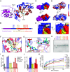
Bisulfite-sequencing reveals a single m42C residue in the 16S rRNA helix 31 loop and the enzyme responsible for its installation. (A) Bisulfite-sequenced RNA collected from TS559 or ΔTK2045 and visualized using Integrative Genomics Viewer where all cytidines were replaced with thymines in the reference genome. Sites of modification are indicated where a cytidine is retained (red) within a sequenced read. Coordinate genome positions in wild-type strain KOD1 and 16S rRNA positions are listed. (B) Purification schematic to isolate the H31-containing fragment from ribosomal or total RNA. (C) The supernatant (wash) and streptavidin capture fractions were analyzed on a polyacrylamide gel. Only when the biotinylated DNA oligo was included was the H31-containing fragment purified (arrow). (D) LC-MS/MS analysis of H31 fragments either derived from the cell or synthesized by in vitro transcription (IVT). (E) The nucleoside structure of m42C. (F) Sequence and modification profile of the H31 region. The colors of subfragments are consistent with those illustrated in panel D. The H31 sequence is underlined. (G) Cryo-EM structure of H31 nucleotides in the T. kodakarensis 70S ribosome. (H) Genomic deletion of gene TK2045 was confirmed using whole genome sequencing (DNA-seq) and RNA-bisulfite sequencing (RNA-seq). Black lines that connect gray bars represent single reads with a gapped alignment. (I) The frequency of cytidine retention in TS559 (n = 3 replicates) and ΔTK2045 (n = 2 replicates) at position C918 in the 16S rRNA during exponential (expo.) or stationary (stat.) growth phase.
Fig. 2.
Fig. 2.
Mature ribosomes are the identified substrate of m42C synthase. (A) SDS-PAGE of recombinant m42C synthase (rTK2045, ~34 KDa). M = marker. (B) The Cryo-EM resolved structure of the T. kodakarensis 70S ribosome, inclusive of rRNA (16S in blue, 23S in magenta, 5S in black) and ribosomal proteins (green). SDS-PAGE (protein) and agarose gel electrophoresis (rRNA) analysis of ribosomes purified from the parent TS559 and ΔTK2045 strains. (C) In vitro methyltransferase radiolabel assay schematic. (D) SAM-dependent methyltransferase activity of rTK2045 on ribosomes and rRNA purified from TS559 or ΔTK2045. n = 3 replicates. **P = 0.0014. (E and F) Mass spectrometry analysis of H31 fragments derived from in vitro methylated ribosomes. (G and H) Site-specific analysis C918 in H31 fragments methylated in vitro. (I and J) Site-specific analysis m4C918 in H31 fragments methylated in vitro.
Fig. 3.
Fig. 3.
Structural and enzymatic characterization of m42C synthase. (A) Atomic structure of m42C synthase at 2 Å. The N and C termini, α-helices, β-sheets, and SAM molecule are labeled. (B) Linear representation of m42C synthase. The N-terminal domain (magenta), C-terminal domain (blue, red, and yellow), methyltransferase domain (yellow and red), and SAM binding residues (yellow) are labeled. Amino acid residues of interest are annotated. (C) Surface structure (D) SAM and substrate binding pocket, and (E) electrostatic potential of m42C synthase. (F) Cytidine and SAM docked into the active site. (G) Proposed interaction between C918 and the DPPR motif. (H) Western blot analysis of recombinant m42C synthase and its variants. (I and J) Methyltransferase activity of each enzyme variant relative to wild-type protein. WT = wild type, ns = not significant, *P-value ≤ 0.05. (K) Methyltransferase activity over time of each enzyme variant. NE = no enzyme. n = 3 replicates.
Fig. 4.
Fig. 4.
Chemical modifications decorate H31 across domains and are fitness relevant in T. kodakarensis. (A) Sequence alignment of the H31 region across domains (Eu. = Eukarya, Ar. = Archaea, Ba. = Bacteria). Blue and green highlighted nucleotides correspond to H31 stem and loop, respectively. C918 in T. kodakarensis and the equivalent positions in other species are boxed. The coordinate position of the 3'-most aligned nucleotide is listed. Stars indicate conserved nucleotides within the alignment. (B) Modifications to H31 across domains. (C) The interface between H31 nucleotides and the anticodon loop of the P-site tRNA. The E-, P-, and A-site tRNAs are shown in blue, gray, and brown, respectively. The translated mRNA is represented in yellow, and the H31 nucleotides are green and blue, consistent with the color scheme in panel A. (D) Head-to-head growth competition between the parent strain TS559, ΔTK2045, and the D205A mutant strain. CI are represented by ± 1 SE. n = 5 replicates.

References

    1. Martinez-Miguel V. E., et al. , Increased fidelity of protein synthesis extends lifespan. Cell Metab. 33, 2288–2300.e12 (2021). - PMC - PubMed
    1. Evans C. R., Fan Y., Ling J., Increased mistranslation protects E. coli from protein misfolding stress due to activation of a RpoS-dependent heat shock response. FEBS Lett. 593, 3220–3227 (2019). - PMC - PubMed
    1. Orita I., et al. , Random mutagenesis of a hyperthermophilic archaeon identified tRNA modifications associated with cellular hyperthermotolerance. Nucleic Acids Res. 47, 1964–1976 (2019). - PMC - PubMed
    1. Shigi N., et al. , Temperature-dependent biosynthesis of 2-thioribothymidine of Thermus thermophilus tRNA. J. Biol. Chem. 281, 2104–2113 (2006). - PubMed
    1. Droogmans L., et al. , Cloning and characterization of tRNA (m1A58) methyltransferase (TrmI) from Thermus thermophilus HB27, a protein required for cell growth at extreme temperatures. Nucleic Acids Res. 31, 2148–2156 (2003). - PMC - PubMed

LinkOut - more resources