. 2024 Dec 24;8(24):6297-6307.
doi: 10.1182/bloodadvances.2024013327.

Epigenetic features support the diagnosis of B-cell prolymphocytic leukemia and identify 2 clinicobiological subtypes

Affiliations

Epigenetic features support the diagnosis of B-cell prolymphocytic leukemia and identify 2 clinicobiological subtypes

Stella Charalampopoulou et al. Blood Adv. .

Abstract

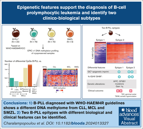
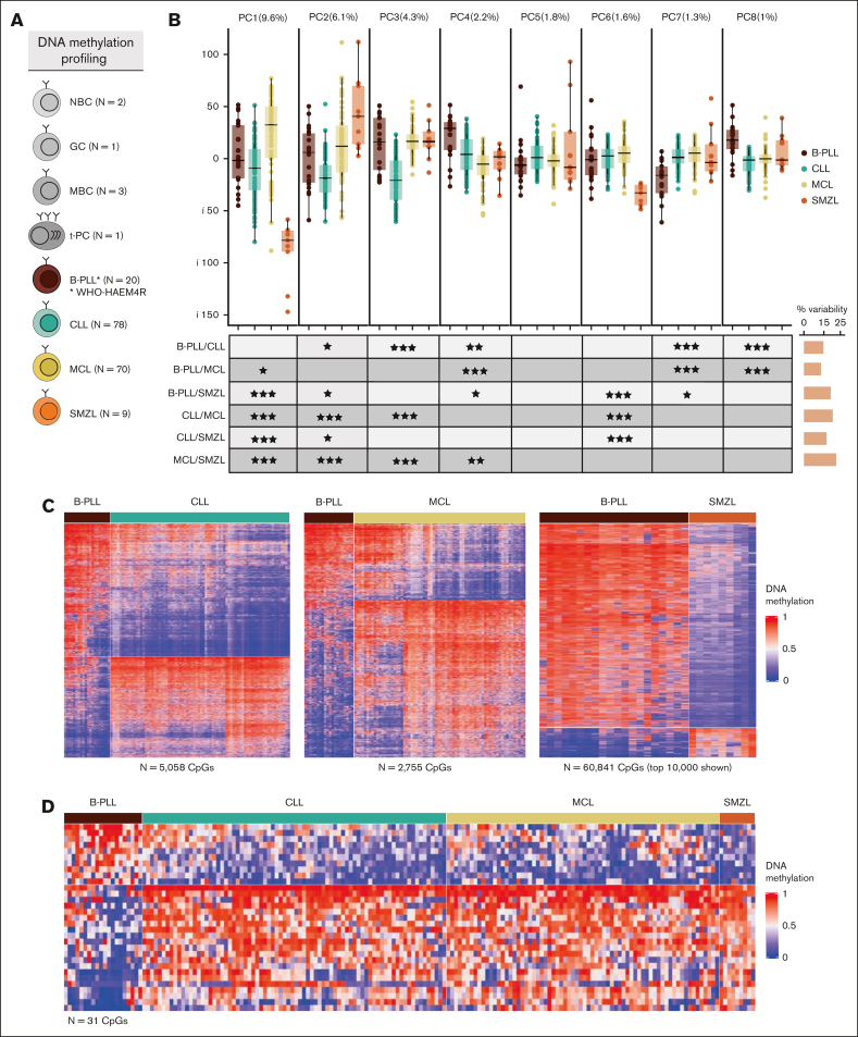
The recognition of B-cell prolymphocytic leukemia (B-PLL) as a separate entity is controversial based on the current classification systems. Here, we analyzed the DNA methylome of a cohort of 20 B-PLL cases diagnosed according to the guidelines of the International Consensus Classification/Fourth revised edition of the World Health Organization Classification, and compared them with chronic lymphocytic leukemia (CLL), mantle cell lymphoma (MCL), splenic marginal zone lymphoma (SMZL), and normal B-cell subpopulations. Unsupervised principal component analyses suggest that B-PLL is epigenetically distinct from CLL, MCL, and SMZL, which is further supported by robust differential methylation signatures in B-PLL. We also observe that B-PLL can be segregated into 2 epitypes with differential clinicobiological characteristics. B-PLL epitype 1 carries lower immunoglobulin heavy variable somatic hypermutation and a less profound germinal center-related DNA methylation imprint than epitype 2. Furthermore, epitype 1 is significantly enriched in mutations affecting MYC and SF3B1, and displays DNA hypomethylation and gene upregulation signatures enriched in MYC targets. Despite the low sample size, patients from epitype 1 have an inferior overall survival than those of epitype 2. This study provides relevant insights into the biology and differential diagnosis of B-PLL, and potentially identifies 2 subgroups with distinct biological and clinical features.


Conflict of interest statement

Conflict-of-interest disclosure: The authors declare no competing financial interests.

Figures

None
Graphical abstract
Figure 1.
Figure 1.
Differential DNA methylation patterns of B-PLL compared with CLL, MCL, and SMZL. (A) Number of samples with DNA methylation data available. (B) Boxplots of the first 8 PCAs including B-PLL, CLL, MCL, and SMZL cases. At bottom, the table shows the false discovery rate (FDR)–corrected P values of Wilcoxon test of pair-wise comparisons across entities. On the right, the bars represent the total percentage of the variability showing significant differences for each PCA comparison. (C) Heat maps of the differentially methylated CpGs between B-PLL and CLL (n = 5058), B-PLL and MCL (n = 2755), and B-PLL and SMZL (n = 60 841). In the case of SMZL, the top 10 000 CpGs with the highest standard deviation are represented. (D) Heat map of the 31 simultaneous differentially methylated CpGs comparing B-PLL to CLL, MCL, and SMZL. FDR levels of significance: FDR < 0.001 = ∗∗∗, FDR < 0.01 = ∗∗, and FDR < 0.05 = ∗.
Figure 2.
Figure 2.
Differential DNA methylation patterns of B-PLL compared with CLL/PL and MCL P/B. (A) Heat map of the differentially methylated CpGs between B-PLL and CLL/PL (n = 3197). (B) PCA of the differentially methylated CpGs between B-PLL and CLL with <10% prolymphocytoid cells (n = 2906), including also the CLL/PL (showing the percentage of cells with prolymphocytoid morphology) and the paired CLL/pPL case. The percentage of prolymphocytes of each of the CLL/PL samples is indicated. (C) Heat map of the differentially methylated CpGs between B-PLL and MCL P/B (n = 10 922). (D) PCA of the differentially methylated CpGs between B-PLL and C1 MCL with classic morphology (n = 8809) including in the plot the C1 MCL P/B.
Figure 3.
Figure 3.
Identification of 2 B-PLL epitypes with signatures related to normal B-cell differentiation. (A) PCA analysis showing PC1 and PC2 with B-PLL and normal B-cell samples. On its side, the boxplots display differences between B-PLL epitypes and normal B cells. (B) Heat map of the differentially methylated CpGs (n = 5465) between B-PLL epitypes. (C) Heat map of the differentially methylated CpGs between B-PLL epitypes related to methylation patterns of NBCs and MBCs (n = 379, of which 249 are hypermethylated and 130 hypomethylated in epitype 1). (D) Percentage of IGHV gene identity B-PLL epitypes. (E) Magnitude of the epiCMIT scores between B-PLL epitypes.
Figure 4.
Figure 4.
Epigenetic and genetic characterization of the differential signature of the B-PLL epitypes. (A) Heat map of the de novo epitype 1 specific methylation signature (n = 799 CpGs). (B) Oncoplot showing genetic alterations of B-PLL cases. (C) Heateukemic nonnodal map of 97 differentially expressed genes between B-PLL epitypes. (D) Gene set enrichment analysis (GSEA) showing enrichment of MYC targets in epitype 1 B-PLL (E) MYC expression percentile of B-PLL, CLL, and MCL cases (F) Bar plot of the numbers of differentially methylated CpGs between CD5+ epitype 2 and CD5 epitype 2 (top) and heat map of DM CpGs between CD5+ epitype 1 and CD5+ epitype 2 (n = 2014; bottom) (G) Kaplan-Meier curves for overall survival between B-PLL epitypes.

References

    1. Algrin C, Pérol L, Chapiro E, et al. Retrospective analysis of a cohort of 41 de novo B-cell prolymphocytic leukemia patients: impact of genetics and targeted therapies (a FILO study) Haematologica. 2023;108(6):1691–1696. - PMC - PubMed
    1. Catovsky D, Galetto J, Okos A, et al. Prolymphocytic leukaemia of B and T cell type. Lancet. 1973;(2):233–234. - PubMed
    1. Galton DAG, Goldman JM, Wiltshaw E, Catovsky D, Henry K, Goldenberg GJ. Prolymphocytic leukaemia. Br J Haematol. 1974;27(1):7–23. - PubMed
    1. Catovsky D, Montserrat E, Muller-Hermelink HK, Harris NL. In: WHO Classification of Tumours - Pathology and Genetics of Tumours of Haematopoietic and Lymphoid Tissue. Jaffe ES, Harris NL, Stein H, Vardiman JW, editors. IARC Press; 2001. B-cell prolymphocytic leukemia; pp. 131–132.
    1. Chapiro E, Pramil E, Diop M, et al. Genetic characterization of B-cell prolymphocytic leukemia: a prognostic model involving MYC and TP53. Blood. 2019;134(21):1821–1831. - PubMed

MeSH terms