. 2024 Dec:78:103400.
doi: 10.1016/j.redox.2024.103400. Epub 2024 Oct 16.

LGR6 protects against myocardial ischemia-reperfusion injury via suppressing necroptosis

Affiliations

LGR6 protects against myocardial ischemia-reperfusion injury via suppressing necroptosis

Mengmeng Zhao et al. Redox Biol. 2024 Dec.

Abstract

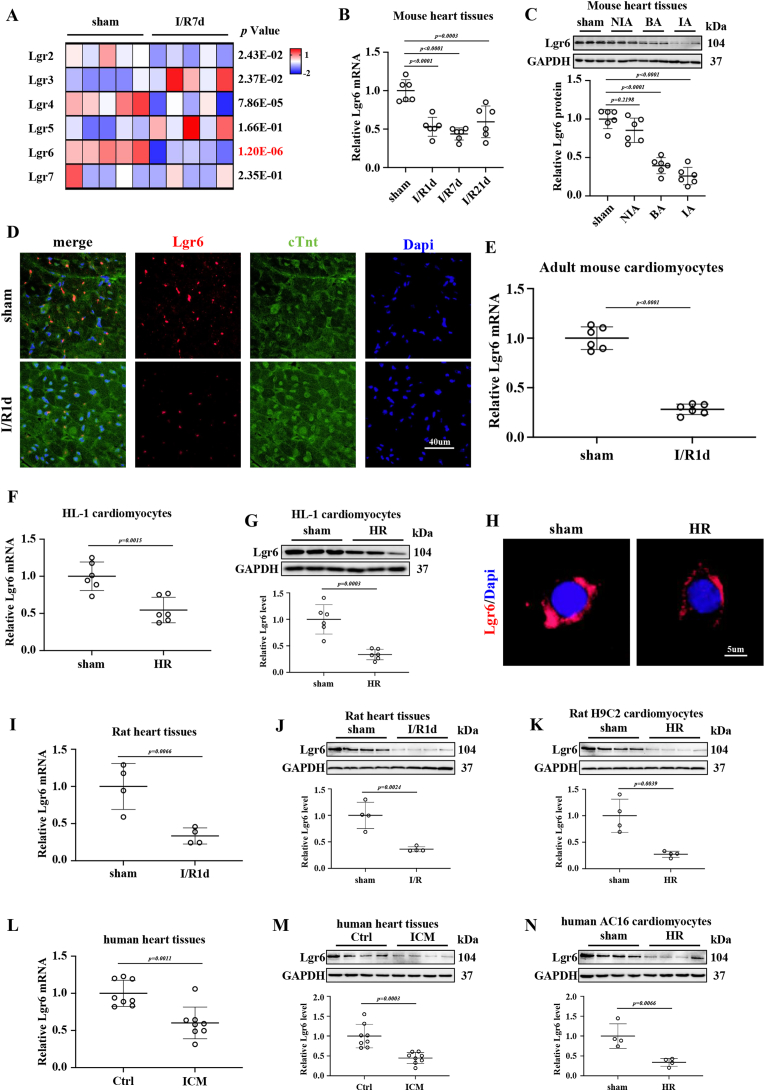
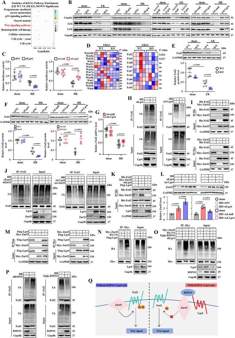
Regulated necrosis (necroptosis) and apoptosis are important biological features of ischemia-reperfusion (I/R) injury. However, the molecular mechanisms underlying myocardial necroptosis remain elusive. Leucine rich repeat containing G protein-coupled receptor 6 (LGR6) has been reported to play important roles in various cardiovascular disease. In this study, we aimed to determine whether LGR6 suppresses I/R-induced myocardial necroptosis and the underlying molecular mechanisms. We generated LGR6 knockout mice and used ligation of left anterior descending coronary artery to produce an in vivo I/R model. The effects of LGR6 and its downstream molecules were subsequently identified using RNA sequencing and CHIP assays. We observed significantly downregulated LGR6 expression in hearts post myocardial I/R and cardiomyocytes post hypoxia and reoxygenation (HR). LGR6 deficiency promoted and LGR6 overexpression inhibited necroptosis and acute myocardial injury after I/R. Mechanistically, in vivo and in vitro experiments suggest that LGR6 regulates the expression of STAT2 and ZBP1 by activating the Wnt signaling pathway, thereby inhibiting cardiomyocyte necroptosis after HR. Inhibiting STAT2 and ZBP1 effectively alleviated the aggravating effect of LGR6 deficiency on myocardial necroptosis after I/R. Furthermore, activating LGR6 with RSPO3 also effectively protected mice from acute myocardial I/R injury. Our findings reveal that RSPO3-LGR6 axis downregulates the expression of STAT2 and ZBP1 through the Wnt signaling pathway, thereby inhibiting I/R-induced myocardial injury and necroptosis. Targeting the RSPO3-LGR6 axis may be a potential therapeutic strategy to treat myocardial I/R injury.

Keywords: LGR6; Myocardial ischemia-reperfusion injury; Necroptosis; STAT2; ZBP1.


Conflict of interest statement

Declaration of competing interest The authors declare that they have no known competing financial interests or personal relationships that could have appeared to influence the work reported in this paper.

Figures

Image 1
Graphical abstract
Fig. 1
Fig. 1
LGR6 expression is markedly downregulated during myocardial I/R injury. (A) Heatmap showing the cardiac expression of LGR2-7 in sham and I/R mice (n = 5). (B) mRNA levels of cardiac LGR6 on day 1, 7 and 21 post I/R (n = 6). (C) Representative immunoblots and corresponding quantification showing cardiac LGR6 in non-infarct area, border area and infarct area at 1 day post I/R (n = 6). (D) Representative immunofluorescence staining images of cardiac tissues for troponin and LGR6 (n = 4). (E) mRNA levels of LGR6 in adult mice cardiomyocytes at 1 day post I/R (n = 6). (F) mRNA levels of LGR6 in HL1 mouse cardiomyocytes (n = 6). (G) Representative immunoblots and corresponding quantification showing LGR6 in HL1s (n = 6). (H) Representative immunofluorescence staining images for LGR6 in HL1s (n = 4). (I) mRNA levels of cardiac LGR6 in rats at 1 day post I/R (n = 4). (J) Representative immunoblots and corresponding quantification showing cardiac LGR6 in border area of rats at 1day post I/R (n = 4). (K) Representative immunoblots and corresponding quantification showing LGR6 in H9C2 rat cardiomyocytes (n = 4). (L) mRNA levels of cardiac LGR6 of patients with normal myocardium (Ctrl) and patients with ischemic cardiomyopathy (ICM) (n = 8). (M) Representative immunoblots and corresponding quantification showing cardiac LGR6 (n = 8). (N) Representative immunoblots and corresponding quantification showing LGR6 in AC16 human cardiomyocytes (n = 4). LGR6, G protein-coupled receptor containing leucine-rich repeats 6; NIA, non-infarct area; BA, border area; IA, infarct area; TNI, troponin. HR, hypoxia and reoxygenation. The data are shown as the mean ± SD. B and C were analyzed by 1-way ANOVA and Tukey's post hoc test. A, E, F, G, I, J, K, L, M and N were analyzed by two-tailed unpaired Student's t-test. p < 0.05 was considered significant.
Fig. 2
Fig. 2
LGR6 knockout accelerates cardiomyocyte necroptosis and acute myocardial injury post I/R. (A) Representative Evans blue and TTC-stained heart sections from global LGR6 KO mice and WT littermates, and the relative ratios of infarct area (IA, pale) to left ventricular were compared between LGR6 KO and WT hearts 24 h after I/R surgery (n = 6). (B) Serum levels of cTNT, CK-MB and LDH (n = 6). (C–H) Representative images and corresponding quantification for vacuolated cardiomyocytes, reactive oxygen species (ROS), TUNEL and PI (n = 6). (I–L) Representative images for c-Cas3, p-Mlkl, p-Ripk1, p-Ripk3 and cTNT (n = 4). (M) Representative immunoblots for p-Mlkl, p-Ripk1 and p-Ripk3 (n = 6). c-Cas3, cleaved-caspase3; p-Mlkl, phosphorylated mixed-lineage kinase domain-like protein; p-Ripk1, phosphorylated receptor-interacting protein kinase 1; p-Ripk3, phosphorylated receptor-interacting protein kinase 3; cTNT, cardiac troponin T; CK-MB, creatine kinase, MB form; LDH, lactate dehydrogenase. The data are shown as the mean ± SD. A was analyzed by two-tailed unpaired Student's t-test. B, D, E, F and G were analyzed by 2-way ANOVA and Tukey's post hoc test. p < 0.05 was considered significant. (For interpretation of the references to colour in this figure legend, the reader is referred to the Web version of this article.)
Fig. 3
Fig. 3
Cardiomyocyte-specific LGR6 overexpression ameliorates acute myocardial injury post I/R. (A) Representative Evans blue and TTC-stained heart sections from mice with AAV9-Lgr6 and AAV9-null, and the relative ratios of infarct area (IA, pale) to left ventricular were compared 24 h after I/R surgery (n = 6). (B) Serum levels of cTNT, CK-MB and LDH (n = 6). (C–E) Representative images and corresponding quantification for vacuolated cardiomyocytes and reactive oxygen species (ROS) (n = 6). (F–J) Representative images and corresponding quantification for PI, p-Mlkl, p-Ripk1, p-Ripk3 and cTNT (n = 4). (K) Representative immunoblots and corresponding quantification for p-Mlkl, p-Ripk1 and p-Ripk3 (n = 6). p-Mlkl, phosphorylated mixed-lineage kinase domain-like protein; p-Ripk1, phosphorylated receptor-interacting protein kinase 1; p-Ripk3, phosphorylated receptor-interacting protein kinase 3; cTNT, cardiac troponin T; CK-MB, creatine kinase, MB form; LDH, lactate dehydrogenase. The data are shown as the mean ± SD. A was analyzed by two-tailed unpaired Student's t-test. B, D, E, J and K were analyzed by 2-way ANOVA and Tukey's post hoc test. p < 0.05 was considered significant. (For interpretation of the references to colour in this figure legend, the reader is referred to the Web version of this article.)
Fig. 4
Fig. 4
RSPO3-LGR6 axis activates Wnt signaling post myocardial I/R. (A) KEGG pathway enrichment of downregulated genes. (B) Representative immunoblots for Ctnnb1, Tcf1 and Lef1 (n = 4). (C) TOPflash reporter assays showing relative luciferase activity (n = 6). (D) Heatmap of genes for Wnt ligands and FZD receptors (n = 4). (E) Protein levels for cardiac Fzd2 (n = 6). (F–G) Protein and mRNA levels for Fzd2 in HL1 cardiomyocytes (n = 6). (H) Representative immunoblots showing ubiquitination of Fzd2 (n = 4). (I) HEK293T cells were transfected for 24 h with plasmids encoding either His-Fzd2 or Myc-Znrf3 alone or in combination. Cell lysates were immunoprecipitated with His and Myc antibodies, and immunoblotting was performed using His or Myc antibodies. (J) Representative immunoblots showing ubiquitination of Fzd2 (n = 4). (K) HEK293T cells were transfected for 24 h with plasmids encoding His-Fzd2, Myc-Znrf3, sh-Lgr6 and Flag-Lgr6 to examine the Znrf3 level bounding to Fzd2. (L) Representative immunoblots and corresponding quantification for Znrf3 (n = 4). (M) HEK293T cells were transfected for 24 h with plasmids encoding either Flag-Lgr6 or Myc-Znrf3 alone or in combination. Cell lysates were immunoprecipitated with Flag and Myc antibodies, and immunoblotting was performed using Flag or Myc antibodies. (N) HEK293T cells were transfected for 24 h with plasmids encoding Flag-Lgr6, Myc-Znrf3 and HA-Ub. Cell lysates were immunoprecipitated with HA antibodies, and immunoblotting was performed using Myc antibodies. (O) HEK293T cells were transfected for 24 h with plasmids encoding Egfp-RSPO3, Myc-Znrf3, sh-Lgr6 and HA-Ub. Cell lysates were immunoprecipitated with Myc antibodies, and immunoblotting was performed using HA antibodies. (P) Representative immunoblots showing ubiquitination of Fzd2 (n = 4). (Q) Proposed mechanisms, RSPO3-LGR6 axis regulates the activation of the Wnt signaling pathway after HR in cardiomyocytes by regulating Fzd2 ubiquitination. RSPO3, R-spondin 3; Lgr6, G protein-coupled receptor containing leucine-rich repeats 6; KEGG, Kyoto Encyclopedia of Genes and Genomes; CTNNB1, catenin beta 1; Tcf1, T-cell factor 1; Lef1, lymphoid enhancer-binding factor 1; Fzd, frizzled; Znrf3, Zinc/RING finger protein 3. The data are shown as the mean ± SD. C, E, F and G were analyzed by 2-way ANOVA and Tukey's post hoc test. L was analyzed by 1-way ANOVA and Tukey's post hoc test. p < 0.05 was considered significant.
Fig. 5
Fig. 5
LGR6 deficiency promoted Stat2 expression in cardiomyocyte post HR. (A) Diagram of workflow for the selection of potential genes regulating necroptosis from the CHIP-seq datasets of Lef1 and differently expressed genes identified in L6KO hearts. (B) CHIP-seq data sets of Lef1. (C) Volcano plot of differently expressed genes in L6KO hearts by comparison with WT hearts 1 day post I/R. (D) Comparison between common genes from CHIP-seq data sets and differently expressed genes identified in L6KO hearts. (E) Heatmap of differently expressed genes in KEGG Necroptosis pathway (n = 4). (F) Representative immunoblots and corresponding quantification for cardiac Stat2 (n = 6). (G) Representative images and corresponding quantification for Stat2 and cTNT (n = 6). (H–I) Representative immunoblots and corresponding quantification for Stat2 in HL1 cardiomyocytes (n = 6 or 4). (J–K) ChIP-qPCR assay in HL1 cells for Lef1 or IgG occupancy at Stat2 promoter fragments (n = 4). (L) Luciferase activation driven by Stat2 promoter after normalization to Renilla luciferase in HEK293T cells (n = 4). (M) Luciferase activation driven by Stat2 promoter after normalization to Renilla luciferase in HL1 cells (n = 4). (N) IGV tracks showing RNA-seq of Stat2 gene (upper), and Lef1 ChIP-Seq signals at Stat2 gene locus (lower). (O) Consensus DNA-binding motifs of Lef1 according to JASPAR database. (P) Schematic diagram of the construction of wild type and mutant luciferase reporter plasmids of Stat2 promoter. (Q) Luciferase activation driven by the wild type or mutant Stat2 promoter after normalization to Renilla luciferase in HEK293T cells (n = 4). The data are shown as the mean ± SD. F and H were analyzed by 2-way ANOVA and Tukey's post hoc test. I, L, M and Q were analyzed by 1-way ANOVA and Tukey's post hoc test. K was analyzed by two-tailed unpaired Student's t-test. p < 0.05 was considered significant.
Fig. 6
Fig. 6
Stat2 activates Zbp1 transcriptional activity. (A) Representative immunoblots and corresponding quantification for cardiac Zbp1 (n = 6). (B) Representative images for Zbp1 and cTNT (n = 6). (C–E) Representative immunoblots and corresponding quantification for ZBP1 in HL1 cardiomyocytes (n = 6 or 4). (F) ChIP-qPCR assay in HL1 cells for Stat2, Lef1 or IgG occupancy at Zbp1 promoter fragments (n = 4). (G) Luciferase activation driven by Zbp1 promoter after normalization to Renilla luciferase in HEK293T cells (n = 4). (H) Luciferase activation driven by Zbp1 promoter after normalization to Renilla luciferase in HL1 cells (n = 4). (I) IGV tracks showing RNA-seq of Zbp1 gene (upper), and Stat2 ChIP-Seq signals at Zbp1 gene locus (lower). (J) Consensus DNA-binding motifs of Stat2 according to JASPAR database. (K) Schematic diagram of the construction of wild type and mutant luciferase reporter plasmids of Zbp1 promoter. (L) Luciferase activation driven by the wild type or mutant Zbp1 promoter after normalization to Renilla luciferase in HEK293T cells (n = 4). The data are shown as the mean ± SD. A and C were analyzed by 2-way ANOVA and Tukey's post hoc test. D, E, F, G, H and L were analyzed by 1-way ANOVA and Tukey's post hoc test. p < 0.05 was considered significant.
Fig. 7
Fig. 7
Cardiomyocyte-specific Stat2 and Zbp1 knockdown abolishes acute myocardial injury and necroptosis post I/R exacerbated by LGR6 deletion. (A) Protocol diagram for the study design. (B) Serum levels of cTNT, CK-MB and LDH (n = 6). (C) Representative images and corresponding quantification for vacuolated cardiomyocytes and ROS (n = 4). (D–H) Representative images and corresponding quantification for PI, p-Mlkl, p-Ripk1, p-Ripk3 and cTNT (n = 4). (I) Representative immunoblots and corresponding quantification for p-Mlkl, p-Ripk1 and p-Ripk3 (n = 4). The data are shown as the mean ± SD. All data were analyzed by 1-way ANOVA and Tukey's post hoc test. p < 0.05 was considered significant.
Fig. 8
Fig. 8
Lgr6 activation via recombinant RSPO3 protected mice from acute myocardial injury post I/R. (A) Protocol diagram for the study design. (B) Serum levels of cTNT in I/R mice with different doses of RSPO3 treatment (n = 6). (C) Representative Evans blue and TTC-stained heart sections, and the relative ratios of infarct area (IA, pale) to left ventricular were compared 24 h after I/R surgery (n = 6). (D–E) Representative images and corresponding quantification for vacuolated cardiomyocytes and ROS (n = 4). (F) Representative images and corresponding quantification for PI and cTNT (n = 4). (G) Representative images for p-Mlkl, p-Ripk1, p-Ripk3 and cTNT (n = 4). (H) Representative immunoblots and corresponding quantification for cardiac p-Mlkl, p-Ripk1, p-Ripk3, Fzd2, Ctnnb1, Lef1, Stat2 and Zbp1 (n = 4). (I) Proposed mechanisms. RSPO3-LGR6 axis inhibited acute myocardial I/R-induced injury and necroptosis by regulating STAT2-ZBP1 signaling pathway. The data are shown as the mean ± SD. B, D, E, F and H were analyzed by 1-way ANOVA and Tukey's post hoc test. C was analyzed by two-tailed unpaired Student's t-test. p < 0.05 was considered significant. (For interpretation of the references to colour in this figure legend, the reader is referred to the Web version of this article.)

References

    1. Algoet M., et al. Myocardial ischemia-reperfusion injury and the influence of inflammation. Trends Cardiovasc. Med. 2023;33:357–366. doi: 10.1016/j.tcm.2022.02.005. - DOI - PubMed
    1. Filipowska J., Kondegowda N.G., Leon-Rivera N., Dhawan S., Vasavada R.C. LGR4, a G Protein-Coupled receptor with a systemic role: from development to metabolic regulation. Front. Endocrinol. 2022;13 doi: 10.3389/fendo.2022.867001. - DOI - PMC - PubMed
    1. Huang C.K., et al. Lgr4 governs a pro-inflammatory Program in macrophages to antagonize post-infarction cardiac repair. Circ. Res. 2020;127:953–973. doi: 10.1161/circresaha.119.315807. - DOI - PubMed
    1. Li H., et al. Maresin 1 intervention reverses experimental pulmonary arterial hypertension in mice. Br. J. Pharmacol. 2022;179:5132–5147. doi: 10.1111/bph.15906. - DOI - PubMed
    1. Elder C.T., et al. Maresin 1 activates LGR6 signaling to inhibit smooth muscle cell activation and attenuate murine abdominal aortic aneurysm formation. Faseb. J. 2021;35 doi: 10.1096/fj.202100484R. - DOI - PMC - PubMed

Publication types

Substances

LinkOut - more resources