. 2024 Nov 7;111(11):2566-2581.
doi: 10.1016/j.ajhg.2024.10.002. Epub 2024 Oct 28.

Homozygous variants in WDR83OS lead to a neurodevelopmental disorder with hypercholanemia

Affiliations

Homozygous variants in WDR83OS lead to a neurodevelopmental disorder with hypercholanemia

Scott Barish et al. Am J Hum Genet. .

Abstract

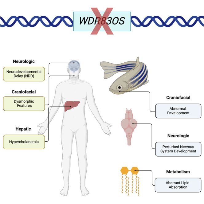
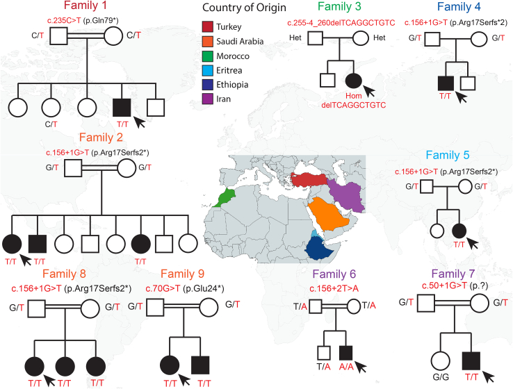
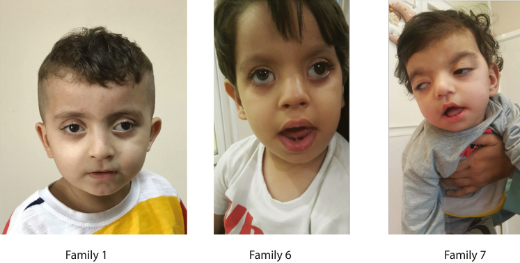
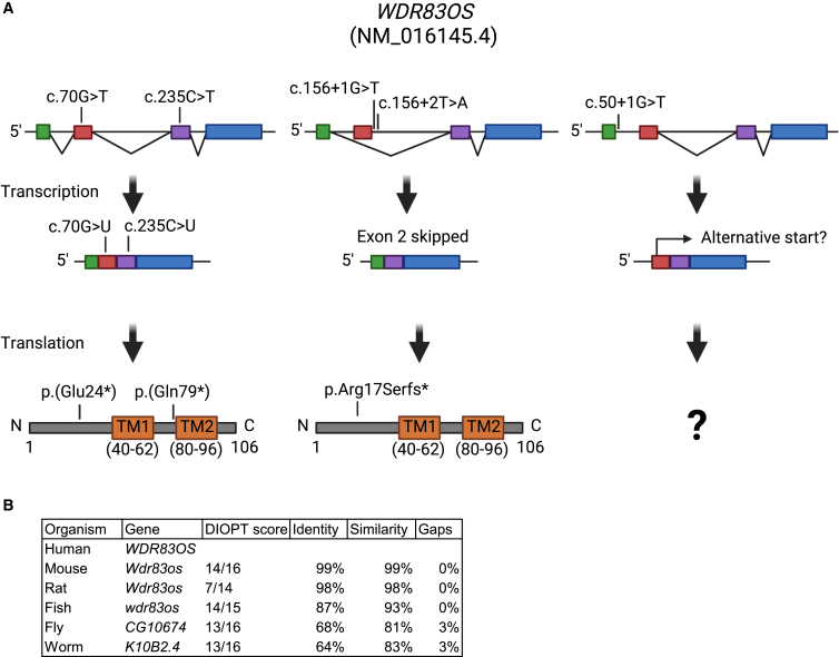
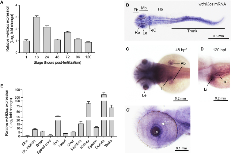
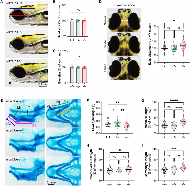
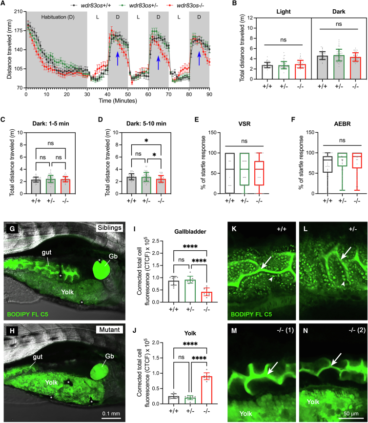
WD repeat domain 83 opposite strand (WDR83OS) encodes the 106-aa (amino acid) protein Asterix, which heterodimerizes with CCDC47 to form the PAT (protein associated with ER translocon) complex. This complex functions as a chaperone for large proteins containing transmembrane domains to ensure proper folding. Until recently, little was known about the role of WDR83OS or CCDC47 in human disease traits. However, biallelic variants in CCDC47 were identified in four unrelated families with trichohepatoneurodevelopmental syndrome, characterized by a neurodevelopmental disorder (NDD) with liver dysfunction. Three affected siblings in an additional family share a homozygous truncating WDR83OS variant and a phenotype of NDD, dysmorphic features, and liver dysfunction. Using family-based rare variant analyses of exome sequencing (ES) data and case matching through GeneMatcher, we describe the clinical phenotypes of 11 additional individuals in eight unrelated families (nine unrelated families, 14 individuals in total) with biallelic putative truncating variants in WDR83OS. Consistent clinical features include NDD (14/14), facial dysmorphism (13/14), intractable itching (9/14), and elevated bile acids (5/6). Whereas bile acids were significantly elevated in 5/6 of individuals tested, bilirubin was normal and liver enzymes were normal to mildly elevated in all 14 individuals. In three of six individuals for whom longitudinal data were available, we observed a progressive reduction in relative head circumference. A zebrafish model lacking Wdr83os function further supports its role in the nervous system, craniofacial development, and lipid absorption. Taken together, our data support a disease-gene association between biallelic loss-of-function of WDR83OS and a neurological disease trait with hypercholanemia.

Keywords: ASTERIX; CCDC47; ER translocation; PAT complex; WDR83OS; developmental delay; hypercholanemia; intellectual disability; pruritus.


Conflict of interest statement

Declaration of interests The Department of Molecular & Human Genetics at Baylor College of Medicine receives revenue from clinical genetic testing conducted at Baylor Genetics Laboratories. J.R.L. serves on the Scientific Advisory Board of Baylor Genetics. J.R.L. has stock ownership in 23andMe, is a paid consultant for Genome International, and is a co-inventor on multiple United States and European patents related to molecular diagnostics for inherited neuropathies, eye diseases, genomic disorders, and bacterial genomic fingerprinting.

Figures

None
Graphical abstract
Figure 1
Figure 1
Pedigrees of families carrying homozygous variants in WDR83OS Coding sequence and predicted protein change are indicated above each pedigree, and genotype is displayed next to each sequenced family member. Affected individuals are indicated by black shapes and initial proband of interest by black arrows. Affected individuals’ country of origin is indicated with different colors and shown on the map. Chromatographs where available for Sanger verification can be found in Figure S1.
Figure 2
Figure 2
Dysmorphic features of WDR83OS-affected individuals Pictures of probands from Families 1 (age 3.5 years), 6 (age 26 months), and 7 (age 17 months). Note strabismus and prominent philtrum in all three individuals, tented upper lip and everted lower lip in Individuals 2 and 3, ptosis in Individuals 1 and 3, hypotelorism in Individuals 1 and 2, and hypertelorism in Individual 3.
Figure 3
Figure 3
WDR83OS variants are predicted to cause loss of function (A) Locations of identified stopgain variants (left), splicing variants at the second exon (middle), and first exon (right). Predicted effects on splicing are indicated by black triangles, and the predicted transcripts and protein products are displayed below. Transmembrane domains (TM1 and TM2) are depicted in orange. The c.156+1G>T variant in the splice donor of exon 2 was shown to cause skipping of exon 2 leading to a premature stop codon. While the c.156+2T>A variant has not been confirmed to cause the same change in the transcript, we expect that the effect of this variant will be similar. The variant in the first exon may cause first-exon skipping and use of an alternative start site. The resulting potential protein would lack both transmembrane domains. Predicted impact of stopgain and frameshift variants at the RNA (transcription) and protein (translation) level are shown in the second and third lines. All three variants would impact one or both the transmembrane domains. (B) Homology of predicted WDR83OS orthologs. Predicted orthologs show high DIOPT, identity, and similarity score, indicating the WDR83OS is well conserved.
Figure 4
Figure 4
Investigation of spatial and temporal expression of wdr83os mRNA (A) The temporal expression of wdr83os mRNA was analyzed by RT-qPCR at different developmental stages of zebrafish. Experiments were performed with biological triplicates and technical triplicates. Expression levels were normalized to the 18S housekeeping gene and compared to the 1 hour post-fertilization (hpf) embryos. Error bar indicates mean ± SD. (B–D) Whole-mount in situ hybridization was conducted on zebrafish at 24, 48, and 120 hpf stages. (B) and (C) are dorsal view; anterior view is to the left. The retina was dissected from (C) and performed in lateral view as shown in (C′). (D) is lateral view, anterior to the left. Fb, forebrain; Mb, midbrain; Hb, hindbrain; Re, retina; Le, lens; TeO, telencephalon; Pb, pectoral fin bud; GCL, ganglion cell layer; Li, liver; Ib, intestinal bulb. (E) The expression of wdr83os mRNA was examined in various adult tissues. Experiments were performed with biological triplicates and technical triplicates. Expression levels were normalized to the 18S housekeeping gene and compared to the skin. Error bar indicates mean ± SD.
Figure 5
Figure 5
Morphological phenotyping of wdr83os mutants showing structural abnormalities (A) Representative image of the wild-type (+/+), heterozygous (+/−) siblings, and homozygous (−/−) wdr83os mutant at 7 dpf. The blue line indicates head size, and the red line indicates eye size. The wdr83os−/− larvae exhibit aberrant lower jaw morphology (black arrowhead) and delayed swim bladder inflation (cyan asterisk). Lateral view, dorsal to the top. (B and C) Quantification of head and eye size, as indicated in (A). +/+, n = 30 larvae; +/− and −/−, n = 32 larvae each. Each dot represents one larva. Error bar indicates mean ± SD. (D) Representative images of normal, wide, and close-set eyes are delineated by two parallel dashed lines. The quantification of the distance between eyes was plotted on the right. Dorsal view, anterior to the top. Scale bar indicates 0.1 mm. Each dot represents one animal. +/+, n = 30 larvae; +/−, n = 34 larvae; −/−, n = 33 larvae. The values are presented using a violin plot as a percentage of the mean value of wild-type siblings. (E) Representative image of Alcian blue-stained wdr83os mutant larvae and siblings at 7 dpf. Mk, Meckel’s cartilage; Pq, palatoquadrate cartilage; Ch, ceratohyal cartilage. Left panel is presented in lateral view and right panel is in ventral view. Scale bar indicates 0.1 mm. (F) Quantification of the lower jaw angle between Meckel’s and palatoquadrate cartilage, as illustrated by magenta lines in (E). (G–I) Quantification of Meckel’s cartilage (G), palatoquadrate cartilage (H), and ceratohyal cartilage (I) length, as indicated by blue, green, and yellow lines in (E), respectively. The values were computed as a percentage of the mean value of wdr83os+/+ siblings and presented using a violin plot. Each dot represents one larva. +/+, n = 22 larvae; +/−, n = 20 larvae; −/−, n = 19 larvae. Error bars indicate mean ± SD. Statistical significance was calculated by Brown-Forsthye and Welch’s ANOVA with Dunnett’s T3 multiple-comparisons test: ns, not significant; p ≥ 0.05, p < 0.05, ∗∗p < 0.01, ∗∗∗p < 0.001, and ∗∗∗∗p < 0.0001.
Figure 6
Figure 6
The wdr83os mutants exhibit gradually declining locomotion and disrupted lipid absorption (A) Locomotor activity analysis. 96-well plates containing 96 larvae at 5 dpf were placed in a recording chamber. Larvae were habituated in the dark for 30 min, followed by three 10-min cycles of alternating light and dark periods. Distance traveled was analyzed. (+/+, n = 39; +/−, n = 56; −/−, n = 40). Error bars indicate mean ± SEM. D, dark period; L, light period; blue arrow marks the 5th minute of darkness. (B) Average distance traveled calculated for each larva during light and dark cycles. Error bars indicate mean ± SD. Each dot represents one larva. (C and D) Distance traveled per 5-min interval. Cumulative distance during each 10-min dark cycle was divided into the first (C) and last (D) 5 min. Error bars indicate mean ± SD. Each dot represents one larva. (E and F) Visual (VSR) and acoustic (AEBR) responses were analyzed in zebrafish larvae at 6 dpf. Responses were calculated as a percentage of the total stimuli and presented as a box-and-whisker plot. Each dot represents one animal. (+/+, n = 40; +/−, n = 55; −/−, n = 40). (G and H) Larvae at 6 dpf were fed BODIPY FL C5 fluorophore. After 4 h, larvae were imaged using confocal microscopy (z stack). Blurry green fluorescence in the gut after z stacking is due to gastrointestinal motility during live imaging. Gb, gallbladder; asterisk, pigment cell. The head is on the right of the images, and the dorsal side is at the top of the images. (I and J) Corrected total cell fluorescence (CTCF) was quantified from the gallbladder (I) and yolk (J) confocal images using ImageJ, normalized to background. n = 3 larvae; each dot represents one selected area within tissue, three areas per tissue. Error bars indicate mean ± SD. (K–N) Representative confocal images of intestines in wdr83os siblings and mutants after BODIPY FL C5 feeding. Arrows highlight the intestinal lumen; arrowheads indicate lipid droplets in intestinal enterocytes. wdr83os−/− mutants showed a lack of intestinal lipid droplets. Statistical analysis was performed by Brown-Forsythe and Welch’s ANOVA with Dunnett’s T3 multiple comparisons test: ns = not significant (p ≥ 0.05), p < 0.05, ∗∗∗∗p < 0.0001.

Similar articles

References

    1. Hawrylycz M.J., Lein E.S., Guillozet-Bongaarts A.L., Shen E.H., Ng L., Miller J.A., van de Lagemaat L.N., Smith K.A., Ebbert A., Riley Z.L., et al. An anatomically comprehensive atlas of the adult human brain transcriptome. Nature. 2012;489:391–399. doi: 10.1038/nature11405. - DOI - PMC - PubMed
    1. Alfadhel M., Umair M., Almuzzaini B., Asiri A., Al Tuwaijri A., Alhamoudi K., Alyafee Y., Al-Owain M. Identification of the TTC26 Splice Variant in a Novel Complex Ciliopathy Syndrome with Biliary, Renal, Neurological, and Skeletal Manifestations. Mol. Syndromol. 2021;12:133–140. doi: 10.1159/000513829. - DOI - PMC - PubMed
    1. Shaheen R., Alsahli S., Ewida N., Alzahrani F., Shamseldin H.E., Patel N., Al Qahtani A., Alhebbi H., Alhashem A., Al-Sheddi T., et al. Biallelic Mutations in Tetratricopeptide Repeat Domain 26 (Intraflagellar Transport 56) Cause Severe Biliary Ciliopathy in Humans. Hepatology. 2020;71:2067–2079. doi: 10.1002/hep.30982. - DOI - PubMed
    1. David O., Eskin-Schwartz M., Ling G., Dolgin V., Kristal E., Benkowitz E., Osyntsov L., Gradstein L., Birk O.S., Loewenthal N., Yerushalmi B. Pituitary stalk interruption syndrome broadens the clinical spectrum of the TTC26 ciliopathy. Clin. Genet. 2020;98:303–307. doi: 10.1111/cge.13805. - DOI - PubMed
    1. Arts H.H., Bongers E.M.H.F., Mans D.A., van Beersum S.E.C., Oud M.M., Bolat E., Spruijt L., Cornelissen E.A.M., Schuurs-Hoeijmakers J.H.M., de Leeuw N., et al. C14ORF179 encoding IFT43 is mutated in Sensenbrenner syndrome. J. Med. Genet. 2011;48:390–395. doi: 10.1136/jmg.2011.088864. - DOI - PubMed

Substances

LinkOut - more resources